Review
. 2009 Oct 14;29(41):12831-8.
doi: 10.1523/JNEUROSCI.3177-09.2009.

Corticostriatal Interactions during Learning, Memory Processing, and Decision Making

Affiliations
Review

Corticostriatal Interactions during Learning, Memory Processing, and Decision Making

Cyriel M A Pennartz et al. J Neurosci. .

Abstract

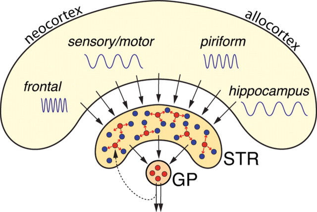
This mini-symposium aims to integrate recent insights from anatomy, behavior, and neurophysiology, highlighting the anatomical organization, behavioral significance, and information-processing mechanisms of corticostriatal interactions. In this summary of topics, which is not meant to provide a comprehensive survey, we will first review the anatomy of corticostriatal circuits, comparing different ways by which "loops" of cortical-basal ganglia circuits communicate. Next, we will address the causal importance and systems-neurophysiological mechanisms of corticostriatal interactions for memory, emphasizing the communication between hippocampus and ventral striatum during contextual conditioning. Furthermore, ensemble recording techniques have been applied to compare information processing in the dorsal and ventral striatum to predictions from reinforcement learning theory. We will next discuss how neural activity develops in corticostriatal areas when habits are learned. Finally, we will evaluate the role of GABAergic interneurons in dynamically transforming cortical inputs into striatal output during learning and decision making.


Figures

Figure 1.
Figure 1.
Schematic representation of frontal hemisections through the rat forebrain and ventral mesencephalon. Axonal projections from the shell target cells in VTA and SNpc that project either (back) to the shell, to the core, or to sensorimotor-related caudate–putamen (blue-green). Fibers originating from the core reach areas in SNpr associated with axonal projections from SNpc to striatal sectors with inputs from anterior cingulate (blue) or prelimbic (purple) cortex. The blue-green region in medial SNpc and dorsal VTA receives inputs from the shell and contains retrogradely labeled cells after tracer injection in sensorimotor striatum. ac, Anterior commissure; SNpr, substantia nigra pars reticulata.
Figure 2.
Figure 2.
Effect of disconnection lesions of the hippocampus (HPC) and nucleus accumbens shell on cue and spatial conditioning. A, Schematic representation of asymmetric, unilateral excitotoxic lesions of the hippocampus and shell. B, Schematic diagram of one chamber of the Y-maze apparatus (only one of the three trays and CS lights are represented here). C, Acquisition of place-cue retrieval following core, shell, disconnection lesions, or disconnection sham operations. The difference score is the number of approaches to CS+ minus the number of approaches to CS−. D, Conditioned place preference performance expressed as the percentage of time spent in each chamber. Data are from Ito et al. (2008).
Figure 3.
Figure 3.
Schematic representation of spontaneous cross-structural replay of place and reward information. A, As a rat runs along a track, hippocampal neurons (HC1–HC4) are activated at specific locations (blue ellipses), whereas a ventral striatal neuron (VS1) is firing before and after reward (R) reception (red ellipse, yellow dot). B, Spike patterns during three task episodes plotted along with hippocampal local field potentials (HC LFP). During posttrack rest periods, replay of firing patterns takes place on an ∼10 times accelerated time scale. Hippocampal neurons are reactivated shortly before the ventral striatal neuron. During pretrack rest, firing patterns were dissimilar to those during track running. See the study by Lansink et al. (2009) for further details.
Figure 4.
Figure 4.
Schematic diagram of parallel cortical–basal ganglia loops contributing to habit formation and maintenance, involving the ventromedial prefrontal cortex (IL) within the limbic–cognitive loop and DLS within the sensorimotor loop. The pseudocolor plot depicts task-related neuronal activity recorded in DLS during training on a T-maze task (modified from Barnes et al., 2005). With overtraining and habit formation, firing patterns develop to accentuate the task start and end (red = greater normalized firing frequency).
Figure 5.
Figure 5.
Striatal microcircuitry in the context of cortical–basal ganglia macrocircuits. Within striatum (STR), FSIs (red) receive complex combinations of cortical inputs and participate in local information processing by influencing nearby MSNs (blue). FSIs also receive a specific feedback input from globus pallidus (GP) that may serve as a more broadly distributed control signal (Gage et al., 2008).

References

    1. Abbott LF, Nelson SB. Synaptic plasticity: taming the beast. Nat Neurosci. 2000;3(Suppl):1178–1183. - PubMed
    1. Aldridge JW, Berridge KC, Rosen AR. Basal ganglia neural mechanisms of natural movement sequences. Can J Physiol Pharmacol. 2004;82:732–739. - PubMed
    1. Alexander GE, Crutcher MD. Functional architecture of basal ganglia circuits: neural substrates of parallel processing. Trends Neurosci. 1990;13:266–271. - PubMed
    1. Alexander GE, Crutcher MD, DeLong MR. Basal ganglia-thalamocortical circuits: parallel substrates for motor, oculomotor, “prefrontal” and “limbic” functions. Prog Brain Res. 1990;85:119–146. - PubMed
    1. Atallah HE, Lopez-Paniagua D, Rudy JW, O'Reilly RC. Separate neural substrates for skill learning and performance in the ventral and dorsal striatum. Nat Neurosci. 2007;10:126–131. - PubMed

Publication types