. 2009 Dec;297(6):R1670-80.
doi: 10.1152/ajpregu.00445.2009. Epub 2009 Oct 14.

Hemorrhagic shock augments lung endothelial cell activation: role of temporal alterations of TLR4 and TLR2

Affiliations

Hemorrhagic shock augments lung endothelial cell activation: role of temporal alterations of TLR4 and TLR2

Yuehua Li et al. Am J Physiol Regul Integr Comp Physiol. 2009 Dec.

Abstract

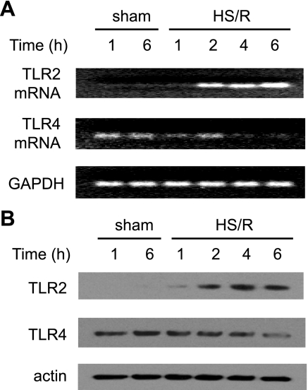
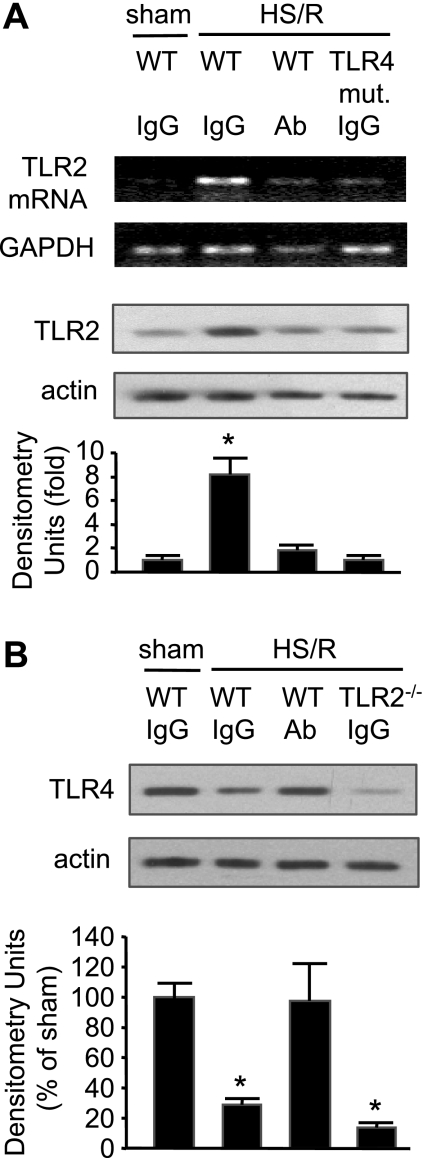
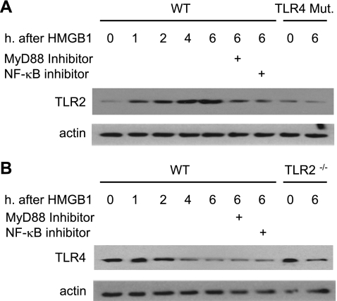
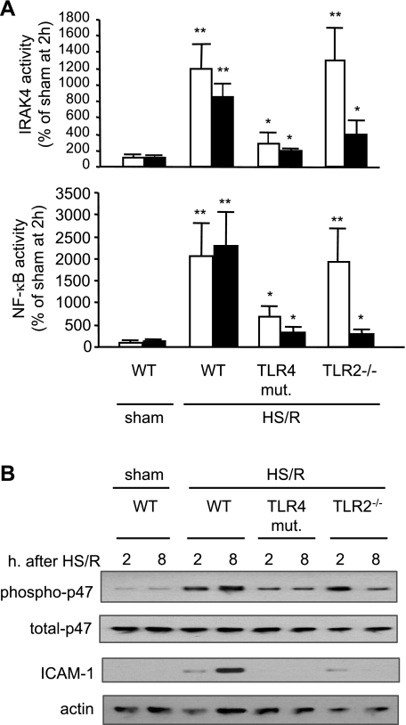
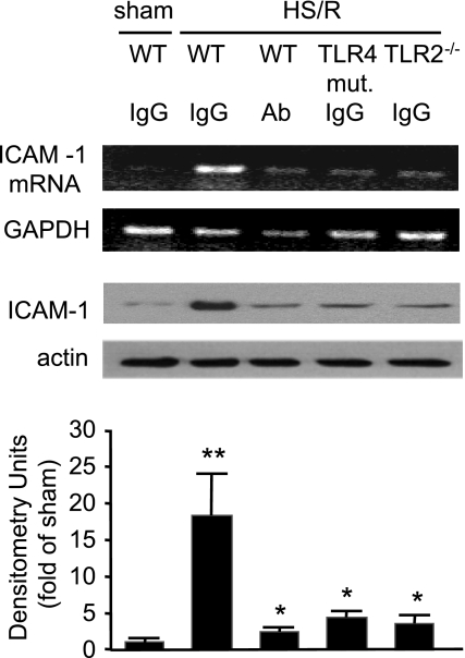
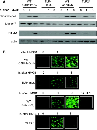
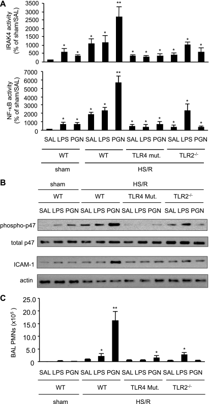
Hemorrhagic shock (HS) due to major trauma predisposes the host to the development of acute lung inflammation and injury. The lung vascular endothelium is an active organ that plays a central role in the development of acute lung injury through generating reactive oxygen species and synthesizing and releasing of a number of inflammatory mediators, including leukocyte adhesion molecules that regulate neutrophils emigration. Previous study from our laboratory has demonstrated that in a setting of sepsis, toll-like receptor-4 (TLR4) signaling can induce TLR2 expression in endothelial cells (ECs), thereby increasing the cells' response to TLR2 ligands. The present study tested the hypothesis that TLR4 activation by HS and the resultant increased TLR2 surface expression in ECs might contribute to the mechanism underlying HS-augmented activation of lung ECs. The results show that high-mobility group box 1 (HMGB1) through TLR4 signaling mediates HS-induced surface expression of TLR2 in the lung and mouse lung vascular endothelial cells (MLVECs). Furthermore, the results demonstrate that HMGB1 induces activation of NAD(P)H oxidase and expression of ICAM-1 in the lung, and MLVECs sequentially depend on TLR4 in the early phase and on TLR2 in the late phase following HS. Finally, the data indicate an important role of the increased TLR2 surface expression in enhancing the activation of MLVECs and augmenting pulmonary neutrophil infiltration in response to TLR2 agonist peptidoglycan. Thus, induction of TLR2 surface expression in lung ECs, induced by HS and mediated by HMGB1/TLR4 signaling, is an important mechanism responsible for endothelial cell-mediated inflammation and organ injury following trauma and hemorrhage.


Figures

Fig. 1.
Fig. 1.
Heat shock resusitation (HS/R) regulation of expression of Toll-like receptor-2 (TLR2) and TLR4 in the lung. Wild-type (WT; C3H/HeOuJ) mice were subjected to HS/R or sham operation. Lung tissue was harvested 1 to 6 h after resuscitation or sham operation, and TLR2 and TLR4 mRNA (A) and protein (B) expression in the lung were detected by RT-PCR and Western blot analysis, respectively. The blots are representative of 3 independent studies; n = 3 mice/group.
Fig. 2.
Fig. 2.
Role of high-mobility group box 1 (HMGB1) in mediating HS/R regulation of TLR2 and TLR4 expression in the lung. A: pretreatment with neutralizing antibody to HMGB1 or TLR4 mutation (mut.) prevents HS/R-induced TLR2 upregulation in the lung. Anti-HMGB1 antibody (Ab; 600 μg per mouse) or nonspecific control IgG was given intraperitoneally to WT (C3H/HeOuJ) or TLR4-mutant mice 10 min before hemorrhage or sham operation. Lung tissue was then collected from the mice at 4 h after HS/R or sham operation for detection of TLR2 mRNA and protein expression using RT-PCR and Western blot analysis, respectively. Images are representative of 3 independent studies. The graph depicts the means ± SE of the fold changes in TLR2 protein expression. *P < 0.01 compared with other groups. B: pretreatment with neutralizing antibody to HMGB1 prevents HS/R-induced TLR4 downregulation in the lung. Anti-HMGB1 antibody or nonspecific control IgG was given to WT (C57BL/6) or TLR2−/− mice 10 min before HS or sham operation, as described in A. Lung tissue was then collected from the mice at 4 h after HS/R or sham operation for detection of TLR4 expression using Western blot analysis. The blots are representative of 3 independent studies. The graph depicts the means ± SE of the %changes in TLR4 expression from 3 mice. *P < 0.01 compared with sham group.
Fig. 3.
Fig. 3.
Dynamic changes in TLR2 and TLR4 in mouse lung vascular endothelial cells (MLVECs) in response to HMGB1. MLVECs were isolated from TLR4-mutant, TLR4 WT (C3H/HeOuJ), TLR2−/−, and TLR2 WT (C57BL/6) mice and incubated with recombinant HMGB1 (0.5 μg/ml) for up to 6 h; TLR2 and TLR4 expression in the MLVECs were then assessed using Western blot analysis. In some experiments, MyD88 inhibitory peptide (100 μM) or NF-κB inhibitor IKK-NBD (100 μM) was added to WT MLVECs 2 h prior to HMGB1. The images are representative of 3 independent studies.
Fig. 4.
Fig. 4.
Time-dependent alteration in TLRs dependency of lung activation following HS/R. WT (C3H/HeOuJ), TLR4-mutant, and TLR2−/− mice were subjected to HS/R or sham operation. Lung tissue was harvested at 2 and 8 h after resuscitation or sham operation, and activities of IRAK4 and nuclear NF-κB (A) as well as phosphorylation of p-47phox and expression of ICAM-1 (B) in the lung were detected. The graph depicts the means ± SE of the %changes, and the white bars and the black bars represent 2 h and 8 h time points, respectively. The graphs and images are representative of 3 independent studies; n = 3 mice/group. *P < 0.01 compared with sham group; **P < 0.01 compared with other groups.
Fig. 5.
Fig. 5.
HS/R-induced ICAM-1 expression in the lung is mediated by HMGB1 through TLR4 and TLR2. TLR4 mutation, TLR2 deficiency, or pretreatment with neutralizing antibody to HMGB1 prevents HS/R-induced ICAM-1 expression in the lung. Anti-HMGB1 antibody (600 μg/mouse ip) or nonspecific control IgG was given intraperitoneally to WT (C3H/HeOuJ), TLR4-mutant, and TLR2−/− mice 10 min before HS/R or sham operation. Lung tissue was then collected from the mice at 8 h after HS/R or sham operation for detection of ICAM-1 expression using RT-PCR and Western blot analysis, respectively. The graph depicts the means ± SE of the %changes in ICAM-1 expression from 3 mice. The images and graphs are representative of at least 3 independent studies. *P < 0.01 compared with sham group; **P < 0.01 compared with other groups.
Fig. 6.
Fig. 6.
TLR4 and TLR2 sequentially mediate HS/R-induced activation of lung ECs. A: MLVECs were isolated from TLR4-mutant, TLR4 WT (C3H/HeOuJ), TLR2−/−, and TLR2 WT (C57BL/6) mice and incubated with recombinant HMGB1 (0.5 μg/ml) for 0 to 8 h; p47phox phosphorylation and ICAM-1 expression in the MLVECs were then assessed. Treatment with HMGB1 for 8 h exhibited a sustained activation of NAD(P)H oxidase and ICAM-1 expression in the WT MLVECs, but not in TLR4-mutant and TLR2−/− MLVECs. B: reactive oxygen species (ROS) production in live MLVECs. MLVECs that were cultured in 12-well cell culture plate were stained with the cell-permeable ROS detection reagent H2DFFDA in the concentration of 10 μM for 10 min. Cells were then washed with HBSS 3 times followed by incubation in the growth medium in the presence or absence of HMGB1 (0.5 μg/ml) for 8 h. The ROS production was then detected by fluorescence microscopy at different time points as indicated. In some experiments, the NAD(P)H oxidase-specific inhibitor diphenyleneiodonium (DPI) (100 μM) was added to the WT (C57BL/6) MLVECs immediately before the treatment of HMGB1 to specify the source of ROS. Blots are representative of at least 3 independent studies.
Fig. 7.
Fig. 7.
Induced TLR2 mediates HS/R-augmented lung activation and inflammation in response to peptidoglycan (PGN). Mice were subjected to HS/R followed by intratracheal PGN (3 μg/kg body wt), LPS (3 μg/kg body wt), or saline (SAL) at 6 h after resuscitation. Activities of IRAK and NF-κB (A), as well as p47phox phosphorylation and ICAM-1 expression (B) in the lung were measured 2 h thereafter. To address the physiological relevance of the altered p47phox phosphorylation and ICAM-1 expression in the lung, PMN in bronchoalveolar lavage fluid were counted 2 h after intratracheal administration of saline, PGN, or LPS (C). The blots and graphs are representative of at least 3 independent studies; n = 3 mice/group. *P < 0.01 compared with control group. **P < 0.01 compared with other groups.

References

    1. Akgur FM, Brown MF, Zibari GB, McDonald JC, Epstein CJ, Ross CR, Granger DN. Role of superoxide in hemorrhagic shock-induced P-selectin expression. Am J Physiol Heart Circ Physiol 279: H791– H797, 2000 - PubMed
    1. Akira S, Takeda K. Toll-like receptor signalling. Nat Rev Immunol 4: 499– 511, 2004 - PubMed
    1. Babior BM. The NADPH oxidase of endothelial cells. IUBMB Life 50: 267– 269, 2000 - PubMed
    1. Bonello S, Zahringer C, BelAiba RS, Djordjevic T, Hess J, Michiels C, Kietzmann T, Gorlach A. Reactive oxygen species activate the HIF-1α promoter via a functional NFκB site. Arterioscler Thromb Vasc Biol 27: 755– 761, 2007 - PubMed
    1. Bowie A, O'Neill LA. The interleukin-1 receptor/Toll-like receptor superfamily: signal generators for pro-inflammatory interleukins and microbial products. J Leukoc Biol 67: 508– 514, 2000 - PubMed

Publication types

MeSH terms

LinkOut - more resources