. 2009 Dec 15;8(24):4091-102.
doi: 10.4161/cc.8.24.10171. Epub 2009 Dec 25.

C. elegans mitotic cyclins have distinct as well as overlapping functions in chromosome segregation

Affiliations

C. elegans mitotic cyclins have distinct as well as overlapping functions in chromosome segregation

Monique van der Voet et al. Cell Cycle. .

Abstract

Mitotic cyclins in association with the Cdk1 protein kinase regulate progression through mitosis in all eukaryotes. Here, we address to what extent mitotic cyclins in the nematode Caenorhabditis elegans provide overlapping functions or distinct biological activities. C. elegans expresses a single A-type cyclin (CYA-1), three typical B-type cyclins (CYB-1, CYB-2.1 and CYB-2.2), and one B3-subfamily member (CYB-3). While we observed clear redundancies between the cyb genes, cyb-1 and cyb-3 also contribute specific essential functions in meiosis and mitosis. CYB-1 and CYB-3 show similar temporal and spatial expression, both cyclins localize prominently to the nucleus, and both associate with CDK-1 and display histone H1 kinase activity in vitro. We demonstrate that inhibition of cyb-1 by RNAi interferes with chromosome congression and causes aneuploidy. In contrast, cyb-3(RNAi) embryos fail to initiate sister chromatid separation. Inhibition of both cyclins simultaneously results in a much earlier and more dramatic arrest. However, only the combination of cyb-1, cyb-3 and cyb-2.1/cyb-2.2 RNAi fully resembles cdk-1 inhibition. This combination of redundant and specific phenotypes supports that in vivo phosphorylation of certain Cdk targets can be achieved by multiple Cdk1/cyclin complexes, while phosphorylation of other targets requires a unique Cdk1/cyclin combination.


Figures

Figure 1
Figure 1
Distinct mitotic cyclin A, B and B3 genes are conserved in Caenorhabditis elegans. (A) Alignment of C. elegans CYA-1, CYB-1, CYB-2.1, CYB-2.2 and CYB-3 coding sequences with ClustalW2 and BoxShade. Asterisks (*) indicate predicted residues of the hydrophobic patch involved in substrate docking. Note that Met144 and Leu191 are not conserved in CYB-3 (B) Phylogenetic tree of the predicted A-, B- and B3-type cyclins from C. elegans (Ce), D. melanogaster (Dm), X. laevis (Xl) and B-type cyclins of S. cerevisiae (Sc) visualized with Phylip DrawTree. Note that the B3-type cyclins are more closely related to cyclin B3 in other species than to other A- and B-type cyclins within the same species.
Figure 2
Figure 2
CYB-1 and CYB-3 bind CDK-1, show associated H1 kinase activity in vitro, and are expressed coincident with cell division. (A) Western blot showing that CYB-1 and CYB-3 antisera are specific. Lanes contain total embryonic protein lysate, obtained after soaking adults in cyb-1 (left), cyb-3 (middle) or lin-5 (right, control) dsRNA. CYB-1 (top), CYB-3 (middle) or anti-α-tubulin antibodies (bottom; loading control) were used for detection. (B) CYB-1 and CYB-3 associate with CDK-1 and (C) form active kinase complexes. Total lysate or immunoprecipitates with the indicated antibodies were used. (D) CYB-1 and CYB-3 protein expression correlate with cell division during development. Protein lysates of synchronized wild-type animals were immunoblotted and probed with the indicated anti-cyclin antibodies. Larval stages are shown. L1 0 hr: developmentally arrested first stage (L1) larvae; L1 10 hr: L1 larvae 10 hr after stimulation of development by food addition. α-Tubulin protein levels serve as a loading control. (E) cyb-1 and cyb-3 RNAi specifically reduce expression of the corresponding proteins. Wild-type or RNAi-treated embryos were triple-stained for DNA (DAPI, left), CYB-1 (middle) and CYB-3 (right). Anterior is to the left, scale bar approx. 10 μm.
Figure 3
Figure 3
CYB-1 and CYB-3 show largely overlapping protein localizations. (A and B) CYB-1 and (C and D) CYB-3 localization are cytoplasmic in meiosis. (E–H) Following the completion of meiosis, CYB-1 and CYB-3 localize to the maternal pronucleus in the anterior (left arrow) and paternal pronucleus (right arrow). CYB-1 in particular is also present in the cytoplasm. (I–L) CYB-1 and CYB-3 remain present in the cytoplasm and nucleus during prophase, but are undetectable in anaphase. Two-cell embryos in which the anterior AB cell is in anaphase (arrowhead) and the posterior P1 cell in prophase (arrow). CYB-1 and CYB-3 are present in prophase but not in anaphase. (M–P) Similar but somewhat later embryos, in which P1 is in metaphase. Embryos were stained for DNA (DAPI) and anti-CYB-1 or CYB-3 antibodies. Anterior is to the left, scale bar approx. 10 μm.
Figure 4
Figure 4
Mitotic defects associated with CYB-1 and CYB-3 knockdown. (A–F) Development of a wild-type embryo from pronuclear formation until the four-cell stage. The metaphase plate is indicated by a single arrow; segregating chromosomes in anaphase by the double arrows. (G–L) cyb-1(RNAi) embryo. Alignment of the chromosomes at the metaphase plate is incomplete; however, chromosome segregation continues, frequently followed by the formation of multiple nuclei within a single cell. Arrows in (K and L) show the formation of multiple nuclei in the posterior P1 cell following the first mitosis and in the EMS cell after division of P1. (C) cyb-3(RNAi) embryo. Black arrows point to metaphase-aligned chromosomes, white arrow point to furrow ingression (in P) and spindle poles (in Q). Sister chromatids fail to separate (note the expanded time in the right image). In most embryos, a cleavage furrow forms, fails to complete abscission and subsequently regresses (D) cyb-1;cyb-3 double RNAi embryo arrests prior to initiation of the first mitosis. A (single) polar body is expelled during meiosis and the pronuclei move together slowly. No further development is observed. Selected images are from time-lapse DIC recordings of living embryos. Scale bar approx. 10 μm. (Y) Timing of events following meiosis, appearance of a maternal pronucleus was taken as t = 0. Wild-type (n = 3), cyb-1 RNAi (n = 3), cyb-3 RNAi (n = 4), cyb-1;cyb-3 RNAi (n = 6). (1) 5/6 cyb-1;cyb-3 RNAi embryos failed pronuclear centration, (2) 5/6 cyb-1;cyb-3 RNAi embryos failed pronuclear rotation, (3) 1/4 cyb-3 RNAi embryos failed cytokinesis.
Figure 5
Figure 5
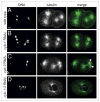
Distinct mitotic defects in cyb-1(RNAi), cyb-3(RNAi) and cyb-1-;cyb-3 double RNAi embryos. Images show similar stage embryos, double-stained for DNA (left) and α-tubulin (middle). Merged images are to the right. (A) wild-type two-cell embryo, with bipolar spindles and AB cell in anaphase (left) and P1 in metaphase (right). (B) two-cell stage cyb-1(RNAi) embryo, arrows indicate unequal DNA segregation in anaphase of AB (see arrow), and a presumed lagging chromosome in metaphase of P1 (right cell, arrow). (C) cyb-3(RNAi) embryo demonstrating lack of chromosome segregation, with continued spindle poles duplication. (D) cyb-1;cyb-3 double RNAi embryo arrested before fusion of the maternal and paternal pronuclei. Anterior is to the left, scale bar 10 μm.
Figure 6
Figure 6
Time-lapse fluorescent microscopy demonstrates meiotic defects. Images show still-shots of meiotic time-lapse movies in utero. (A) wild-type meiosis I and II; chromosomes align at a metaphase spindle (t = 5:30; 19:30), the spindle rotates by 90° (t = 7:30; 23:00), and segregates the chromosomes during anaphase (t = 11.30; 28.00). (B) RNAi of cdk-1. Chromosomes remain in a diakinesis arrangement with limited microtubule organization, the spindle fails to form, but exit from meiosis happens with normal timing. (C) RNAi of cyb-1 results in meiotic defects in chromosome alignment and segregation (white arrow). (D) RNAi of cyb-3 results in a substantial delay in metaphase of meiosis II. (E) Double RNAi of cyb-1 and cyb-3 results in a dramatic meiosis II defect, while meiosis I is delayed but still completes. (F) Triple RNAi of cyb-1, cyb-2.1/2.2, cyb-3 results in diakinesis arrest, similar to cdk-1 RNAi. Red: H2B::Cherry, green: α-tubulin::GFP. Time after entry into the uterus is indicated in each panel. Scale bar is 2 μm, the future anterior cortex is to the left.

Comment in

Similar articles

Cited by

References

    1. Morgan DO. Cyclin-dependent kinases: engines, clocks and microprocessors. Annu Rev Cell Dev Biol. 1997;13:261–91. - PubMed
    1. Satyanarayana A, Kaldis P. Mammalian cell cycle regulation: several Cdks, numerous cyclins and diverse compensatory mechanisms. Oncogene. 2009;28:2925–39. - PubMed
    1. Sherr CJ. Cancer cell cycles. Science. 1996;274:1672–7. - PubMed
    1. Fitch I, Dahmann C, Surana U, Amon A, Nasmyth K, Goetsch L, et al. Characterization of four B-type cyclin genes of the budding yeast Saccharomyces cerevisiae. Mol Biol Cell. 1992;3:805–18. - PMC - PubMed
    1. Richardson H, Lew DJ, Henze M, Sugimoto K, Reed SI. Cyclin-B homologs in Saccharomyces cerevisiae function in S phase and in G2. Genes Dev. 1992;6:2021–34. - PubMed

Publication types

MeSH terms

LinkOut - more resources