

Review
doi: 10.1038/nature08536.
Molecular genetics and imaging technologies for circuit-based neuroanatomy
Affiliations
- PMID: 19829369
- PMCID: PMC2884271
- DOI: 10.1038/nature08536
Item in Clipboard
Review
Molecular genetics and imaging technologies for circuit-based neuroanatomy
Nature.
.
Abstract
Brain function emerges from the morphologies, spatial organization and patterns of connectivity established between diverse sets of neurons. Historically, the notion that neuronal structure predicts function stemmed from classic histological staining and neuronal tracing methods. Recent advances in molecular genetics and imaging technologies have begun to reveal previously unattainable details about patterns of functional circuit connectivity and the subcellular organization of synapses in the living brain. This sophisticated molecular and genetic 'toolbox', coupled with new methods in optical and electron microscopy, provides an expanding array of techniques for probing neural anatomy and function.
Figures

Several viruses display trans-synaptic spread, including pseudorabies virus (PRV) and rabies virus (RV),,,. PRV can be used to differentially trace intact brain circuits by expressing FPs of different colors (A). A drawback of replicating viral vectors such as PRV is that they continue to propagate from cell to cell, making it difficult to discern monosynaptic from polysynaptic patterns of connectivity. An alternative method relies on the generation of recombinant RV deficient for the gene encoding the G-protein coat particle, which is replaced by an EGFP reporter. To target EGFP-expressing RV to desired neuronal subsets, RV particles can be pseudotyped with the EnvA coat protein, which specifically binds TVA class receptors. By targeting neurons for TVA expression, RV infection can thus be limited to only those neurons (B). A clever twist on this approach implemented by Wickersham et al. included the introduction of a plasmid that encodes the wildtype RV G protein, allowing the disarmed EGFP-expressing virus to undergo one round of subsequent infection to presynaptic partners of TVA-targeted neurons. Since only the initially infected neuron contains the wildtype RV G protein, viral spread is halted after one round of monosynaptic jumping. Adding a plasmid encoding a red FP allows the cell originally targeted for infection to be identified amongst the monosynaptic network of GFP labeled cells (C). Green plus signs represent sights of monosynaptic viral transfer; red crosses represent polysynaptic sites that lack viral transfer due to the absence of wildtype G protein.
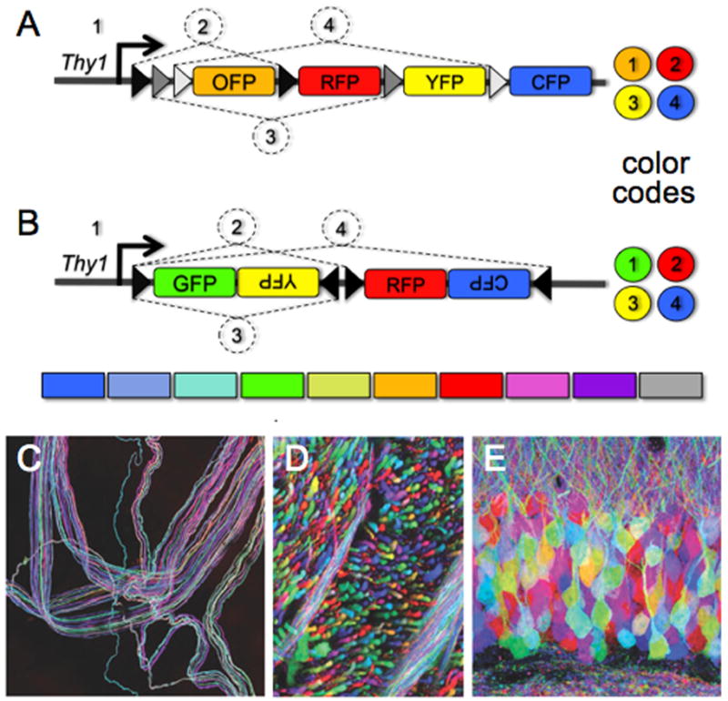
Brainbow mice can be used to identify neuronal subsets by conditional activation of fluorescent reporters. Cre mediated recombination between loxP sequences is orientation dependent. If loxP sites are in the same 5′-3′ direction, recombination results in excision of the flanked DNA sequence. When loxP sites are in opposite orientation, recombination drives inversion of the flanked DNA. A) A Cre-mediated excision strategy based on a transgenic allele containing pairs of mutated loxP sites and multiple fluorescent proteins to generate cells of different colors. Black, gray, and white arrows represent different loxP sequence mutations. Recombination only occurs between mutant pairs, resulting in cells with different color codes depending on the random recombination events between like pairs. The events labeled 1–4 correspond to patterns of FP expression shown on the right. B) A Cre-mediated inversion strategy based on an allele that harbors multiple loxP sites of the same type in both orientations. Stochastic recombination events between inversion pairs results in different color possibilities (1–4, and the resulting color code on right). Bottom, the spectrum of colors that cells can express with different Brainbow alleles and recombination events. C–E) Images of Brainbow tissue following Cre-mediated recombination to stochastically label different types of neurons. Images were originally published in Nat Rev Neurosci, 9, 2008, by J.W. Lichtman, J. Livet, and J.R. Sanes. C) Fibers from a motor nerve innervating the ear. D) Axon tracts coursing through the brainstem. E) Cells of the hippocampal dentate gyrus.
References
-
- Pfister H, Lichtman J, Reid C. The Connectome Project. 2009. < http://iic.harvard.edu/research/connectome>.
-
- Macagno ER, Levinthal C, Sobel I. Three-dimensional computer reconstruction of neurons and neuronal assemblies. Annu Rev Biophys Bioeng. 1979;8:323–351. - PubMed
-
- White JG, Southgate E, Thomson JN, Brenner S. The structure of the nervous system of the nematode Caenorhabditis elegans. Philos Trans R Soc Lond. 1986;314:1–314. - PubMed
-
- Ahmed B, Anderson JC, Martin KA, Nelson JC. Map of the synapses onto layer 4 basket cells of the primary visual cortex of the cat. J Comp Neurol. 1997;380:230–242. - PubMed
Publication types
MeSH terms
Grants and funding
LinkOut - more resources
Full Text Sources
Other Literature Sources
