. 2009 Oct 15;461(7266):947-55.
doi: 10.1038/nature08435.

Asymmetric centrosome inheritance maintains neural progenitors in the neocortex

Affiliations

Asymmetric centrosome inheritance maintains neural progenitors in the neocortex

Xiaoqun Wang et al. Nature. .

Abstract

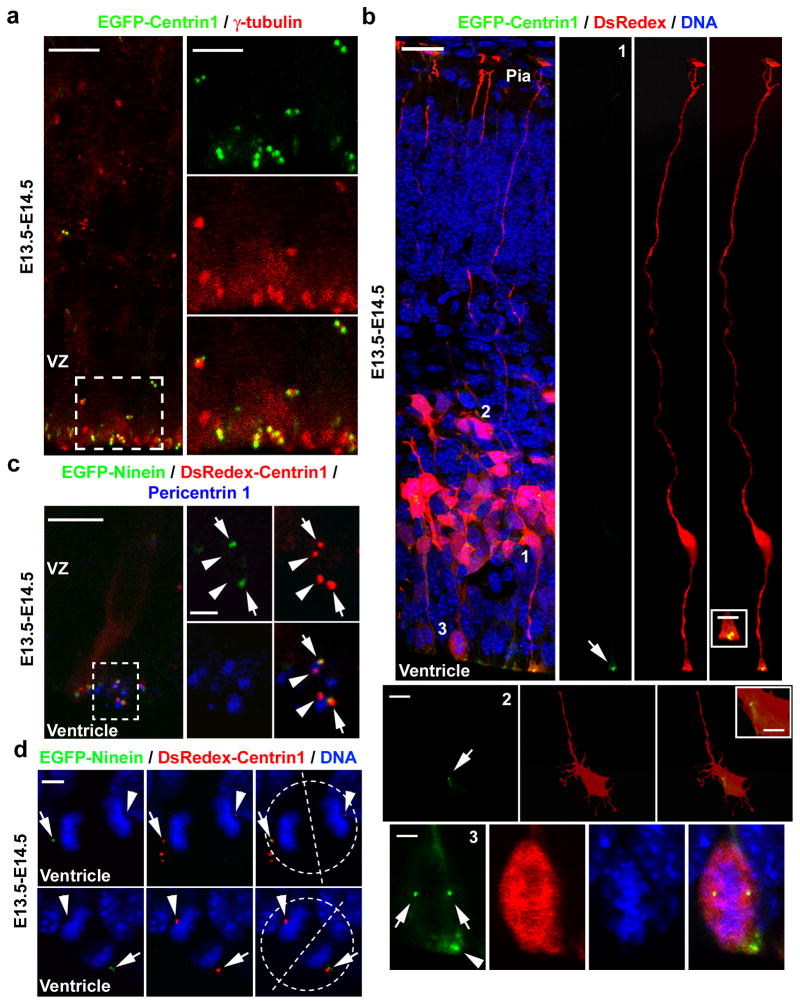
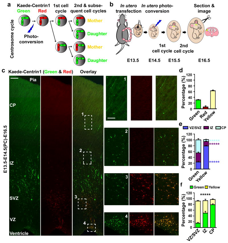
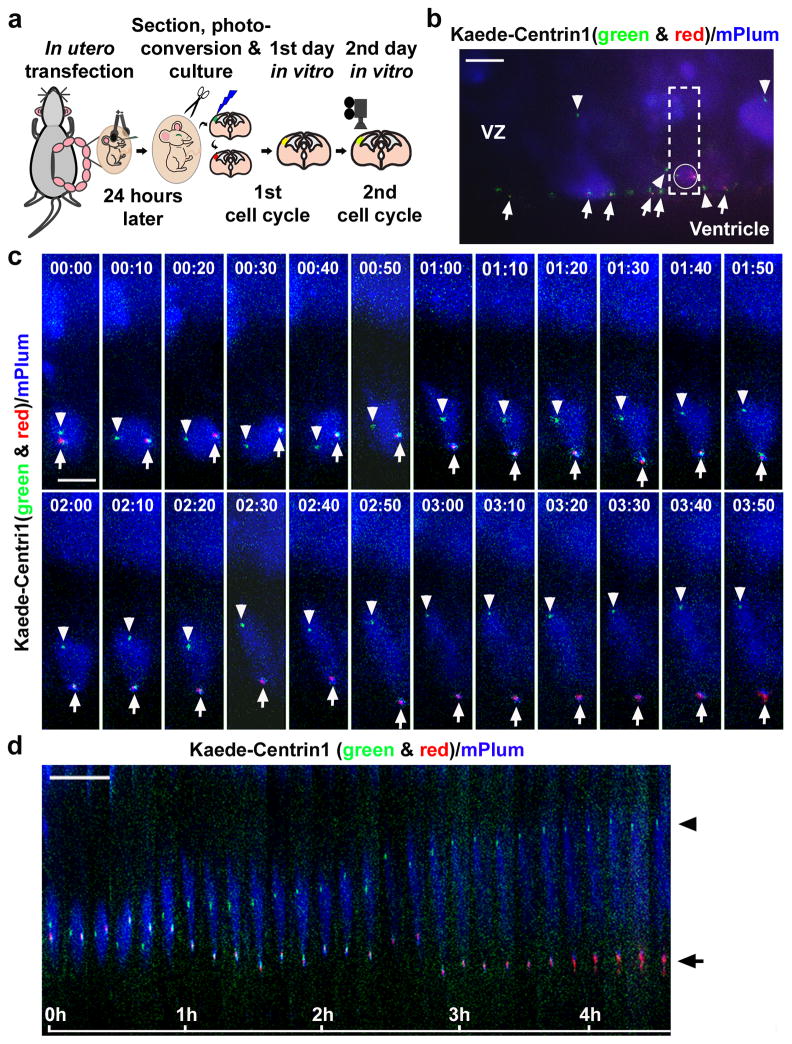
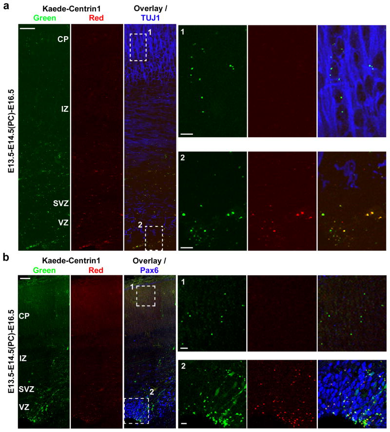
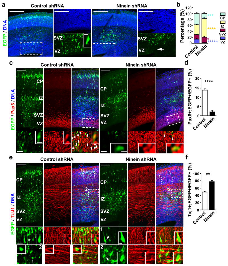
Asymmetric divisions of radial glia progenitors produce self-renewing radial glia and differentiating cells simultaneously in the ventricular zone (VZ) of the developing neocortex. Whereas differentiating cells leave the VZ to constitute the future neocortex, renewing radial glia progenitors stay in the VZ for subsequent divisions. The differential behaviour of progenitors and their differentiating progeny is essential for neocortical development; however, the mechanisms that ensure these behavioural differences are unclear. Here we show that asymmetric centrosome inheritance regulates the differential behaviour of renewing progenitors and their differentiating progeny in the embryonic mouse neocortex. Centrosome duplication in dividing radial glia progenitors generates a pair of centrosomes with differently aged mother centrioles. During peak phases of neurogenesis, the centrosome retaining the old mother centriole stays in the VZ and is preferentially inherited by radial glia progenitors, whereas the centrosome containing the new mother centriole mostly leaves the VZ and is largely associated with differentiating cells. Removal of ninein, a mature centriole-specific protein, disrupts the asymmetric segregation and inheritance of the centrosome and causes premature depletion of progenitors from the VZ. These results indicate that preferential inheritance of the centrosome with the mature older mother centriole is required for maintaining radial glia progenitors in the developing mammalian neocortex.


Figures

Figure 1
Figure 1. Centriole and centrosome asymmetry in the developing neocortex
(a) Images of E14.5 cortices electroporated with EGFP-Centrin1 (green) at E13.5 (E13.5–E14.5) and immunostained for γ-Tubulin (red). (b) Images of cortices electroporated with EGFP-Centrin1 (green) and DsRedexpress (DsRedex, red) and counterstained with DAPI (blue). Arrows and the arrowhead indicate the centrosomes. (c) Images of cortices electroporated with EGFP-Ninein (green, arrows) and DsRedex-Centrin1 (red, arrows and arrowheads) and immunostained for Pericentrin1 (blue). (d) Images of dividing radial glial cells in late mitosis (broken circles) with condensed chromosomes (DAPI, blue) expressing EGFP-Ninein (green, arrows) and DsRedex-Centrin1 (red, arrows and arrowheads). Broken lines indicate the cleavage plane. Scale bars: 10 μm and 5 μm (a); 25 μm, 2.5 μm, 10 μm, 5 μm, and 5 μm (from top to bottom, b); 20 μm and 2 μm (c); 5 μm (d).
Figure 2
Figure 2. Asymmetric segregation of centrosomes with differently aged mother centrioles
(a, b) Strategy and experimental procedure for using Kaede-Centrin1 to distinguish between centrosomes with differently aged mother centrioles. (c) Images of E16.5 cortices electroporated with Kaede-Centrin1 at E13.5 and photo-converted (PC) at E14.5 (E13.5-E14.5(PC)-E16.5). Scale bars: 50 μm and 15 μm. (d–f) Quantifications of the percentage of labelled centrosomes that are green, red, or yellow fluorescent (d), the percentage of green or yellow fluorescent centrosomes that are located in different regions of the developing neocortex (e), and the percentage of labelled centrosomes located in different regions of the developing neocortex that are green or yellow fluorescent (f) (total 4,314 centrosomes from seven individual animals). Data are shown as mean±s.e.m.; *****, p<5e-5.
Figure 3
Figure 3. Distinct behaviour of centrosomes with differently aged mother centrioles
(a) Experimental procedure for time-lapse imaging analysis of centrosome behaviour. (b) Image of a cortical slice in culture expressing Kaede-Centrin1 (green and red) and mPlum (blue) prior to time-lapse imaging. Arrows indicate the centrosomes with the old mother centriole (both green and red fluorescent) and arrowheads indicate the centrosomes with the new mother centriole (green fluorescent only). The outlined region contains a dividing radial glial cell possessing a pair of centrosomes with differently aged mother centrioles (circled). (c, d) Time-lapse (c) and kymograph (d) images of the outlined region in b. The time is indicated at the top (c) or the bottom (d) of images. Arrows indicate centrosomes possessing the old mother centriole and arrowheads indicate centrosomes possessing the new mother centriole. Scale bars: 15 μm (b) and 10 μm (c, d).
Figure 4
Figure 4. Asymmetric inheritance of centrosomes with differently aged mother centrioles
Images of cortices electroporated with Kaede-Centrin1, photo-converted, and immunostained for TUJ1 (a) or for Pax6 (b) (blue). High magnification images of the outlined regions are shown to the right. Scale bars: 50 μm, 10 μm and 10 μm.
Figure 5
Figure 5. Preferential inheritance of the centrosome with the mature mother centriole maintains radial glial progenitors
(a) Images of E16.5 cortices electroporated with either EGFP/Control (left, green) or EGFP/Ninein shRNA (right, green) at E13.5 and counterstained with DAPI (blue). (b) Quantification of the percentage of EGFP-expressing cells in different regions of the developing neocortex (Control shRNA: total 1,873 cells from five individual animals; Ninein shRNA: total 958 cells from five individual animals). (c, e) Images of E16.5 cortices electroporated with EGFP/Control (left, green) or EGFP/Ninein shRNA (right, green) at E13.5 and immunostained for Pax6 (c) or TUJ1 (e) (red). Arrowheads indicate EGFP-expressing cells positive for Pax6 (c). Arrows indicate EGFP-expressing cells that are positive for TUJ1 and the open arrow indicates an EGFP-expressing cell that is not positive for TUJ1 (e). (d, f) Quantification of the percentage of EGFP-expressing cells positive for Pax6 (d; Control shRNA: total 1,752 cells from five individual animals; Ninein shRNA: total 1,230 cells from five animals) or for TUJ1 (f; Control shRNA, total 1,383 cells from five individual animals; Ninein shRNA, total 1,247 cells from five individual animals). Data are shown as mean±s.e.m.; *, p<0.05; **, p<0.005; ***, p<0.001; ****, p<0.0005; *****, p<5e-5. Scale bars: 100 μm, 50 μm, and 10 μm (a); 100μm, 5 μm, and 25 μm (c); 100μm, 25 μm, and 5 μm (e).

Comment in

  • Stem cells: A fateful age gap.
    Stearns T. Stearns T. Nature. 2009 Oct 15;461(7266):891-2. doi: 10.1038/461891a. Nature. 2009. PMID: 19829363 Free PMC article.

References

    1. Miyata T, Kawaguchi A, Okano H, Ogawa M. Asymmetric inheritance of radial glial fibers by cortical neurons. Neuron. 2001;31:727–41. - PubMed
    1. Noctor SC, Flint AC, Weissman TA, Dammerman RS, Kriegstein AR. Neurons derived from radial glial cells establish radial units in neocortex. Nature. 2001;409:714–20. - PubMed
    1. Malatesta P, Hartfuss E, Gotz M. Isolation of radial glial cells by fluorescent-activated cell sorting reveals a neuronal lineage. Development. 2000;127:5253–63. - PubMed
    1. Rakic P. Elusive radial glial cells: historical and evolutionary perspective. Glia. 2003;43:19–32. - PubMed
    1. Miyata T, et al. Asymmetric production of surface-dividing and non-surface-dividing cortical progenitor cells. Development. 2004;131:3133–45. - PubMed

Publication types

MeSH terms