. 2009 Oct 15;4(10):e7482.
doi: 10.1371/journal.pone.0007482.

DUX4c is up-regulated in FSHD. It induces the MYF5 protein and human myoblast proliferation

Affiliations

DUX4c is up-regulated in FSHD. It induces the MYF5 protein and human myoblast proliferation

Eugénie Ansseau et al. PLoS One. .

Abstract

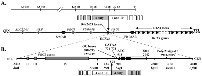
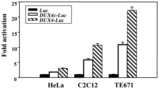
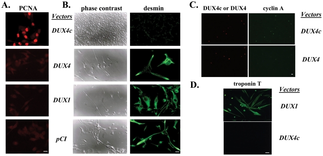
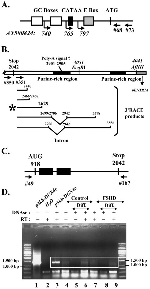
Facioscapulohumeral muscular dystrophy (FSHD) is a dominant disease linked to contractions of the D4Z4 repeat array in 4q35. We have previously identified a double homeobox gene (DUX4) within each D4Z4 unit that encodes a transcription factor expressed in FSHD but not control myoblasts. DUX4 and its target genes contribute to the global dysregulation of gene expression observed in FSHD. We have now characterized the homologous DUX4c gene mapped 42 kb centromeric of the D4Z4 repeat array. It encodes a 47-kDa protein with a double homeodomain identical to DUX4 but divergent in the carboxyl-terminal region. DUX4c was detected in primary myoblast extracts by Western blot with a specific antiserum, and was induced upon differentiation. The protein was increased about 2-fold in FSHD versus control myotubes but reached 2-10-fold induction in FSHD muscle biopsies. We have shown by Western blot and by a DNA-binding assay that DUX4c over-expression induced the MYF5 myogenic regulator and its DNA-binding activity. DUX4c might stabilize the MYF5 protein as we detected their interaction by co-immunoprecipitation. In keeping with the known role of Myf5 in myoblast accumulation during mouse muscle regeneration DUX4c over-expression activated proliferation of human primary myoblasts and inhibited their differentiation. Altogether, these results suggested that DUX4c could be involved in muscle regeneration and that changes in its expression could contribute to the FSHD pathology.


Conflict of interest statement

Competing Interests: The authors have declared that no competing interests exist.

Figures

Figure 1
Figure 1. Localization of the DUX4 and DUX4c genes.
(A) Schematic representation of the 4q35 subtelomeric region with the D4Z4 repeat array and the SLC25A4 (previously known as ANT1) , ALP , FRG1 , TUBB4Q and FRG2 genes. DUX4 maps within each D4Z4 element and DUX4c within an isolated inverted D4Z4 unit at the D4S2463 locus. S/MAR and FR-MAR: nuclear scaffold/matrix attachment regions, . Upper line: 4q35/10q26 limit of homology . (B) Enlargement (inverted orientation) of the 7.5-kb fragment that contains DUX4c with part of the FRG2 gene. The DUX4c ORF is boxed, with the homeoboxes in black. The promoter GC boxes, the putative variant TATA box (CATAA) and polyadenylation signal are indicated. Numbering from the EcoRI site (GenBank acc. no. AY500824).
Figure 2
Figure 2. Transcriptional activity of the DUX4c gene.
Transcriptional activity of the DUX4c promoter. HeLa, C2C12 and TE671 cells were transfected with pGL3 vectors containing the luciferase reporter gene either promoterless (black bars) or fused to the DUX4c (white bars) or DUX4 (striped bars) promoter. Luciferase activity was measured 24 h post-transfection and expressed relative to the activity of the promoterless vector set to 1. The means and standard errors are indicated (n = 18).
Figure 3
Figure 3. DUX4c protein expression in myoblasts.
(A) Transcription/translation in vitro in a rabbit reticulocyte lysate in the presence T7 RNA polymerase and [35S]-cysteine with genomic fragments encoding DUX4 (lane 1) or DUX4c (lane 2) cloned in pCIneo. 52 kDa-DUX4 (white arrow) and 47 kDa-DUX4c (black arrow) are detected by autoradiography after 10% PAGE-SDS. (B–C) 30 µg total proteins extracted from primary myoblast were analysed by 4–12% PAGE-SDS and Western blot with the indicated primary antibodies, appropriate secondary antibodies coupled to HRP and the ECL kit. α-Tubulin was the loading control. (B) Competition: a 5-fold excess of DUX4c antigenic peptide was pre-incubated (+) or not (−) with the serum raised against DUX4c or cadherin as indicated. (C) Extracts were prepared either from proliferating myoblasts or 2 (d2) or 6 (d6) days after induction of differentiation. (D) DUX4c (red) was detected by immunofluorescence in nuclei of myoblasts and myotubes 2 (d2) and 6 (d6) days after inducing differentiation. a' and b' correspond respectively to two enlarged nuclei from d2 and d6 (white boxes in a and b). The labeling is weakened after competition with the immunogenic peptide (c). Troponin T (green) is a myotube differentiation marker. Myoblasts not fused to myotubes express DUX4c (red nuclei) but not troponin T. Bar corresponds to 20 µm. (E) 30 µg protein extracted from primary myotubes were analyzed by Western blot as in (C). (F) Densitometric scanning of the film shown in (E): DUX4c expression levels were normalized to α-Tubulin (relative absorbance units). C: control, F: FSHD, D: DMD.
Figure 4
Figure 4. DUX4c protein expression in muscle biopsies.
(A) 30 µg protein extracted of muscle biopsies were analyzed by Western blot as in Fig. 3, except that cytochrome C was the internal loading control. (B) Densitometric scanning of the Western blot shown in (A) and of additional samples (not shown): DUX4c expression levels were normalized to cytochrome C (relative absorbance units). Samples are indicated C1 to C4 for controls, F1 to F10 for FSHD, and D1 to D4 for DMD as well as the D4Z4 copy numbers of the FSHD patients. The biopsied muscle is indicaded (D, Q: non-affected deltoid or quadriceps; T*: affected trapezius). F10 also has a D4Z4 array contraction on the second 4q35 allele (+7).
Figure 5
Figure 5. DUX4c over-expression induces MYF5.
(A–C) TE671 cells were transfected with the indicated pCIneo vectors. Nuclear extracts were deposited in triplicate on a plate where the MYOD1, MEF2 or MYF5 specific DNA target was immobilized. The DNA-bound protein was detected by ELISA (TransAm assay). Relative absorbances are given relative to the insertless pCIneo sample arbitrarily set to 1. Three independent experiments (1 to 3) made in triplicate are presented. (D) TE671 nuclear extracts were prepared 48 h after transfection as above and 30 or 15 (*) µg were analyzed by 10% PAGE-SDS and Western blotting with a serum raised against MYF5 or actin (internal control). NT: non transfected cells. (E) same as in D but transfection was with pAC1M2-DUX4c and DUX4c expression induced by doxycycline (o to1000 ng). (F) Mouse C2C12 cells were transfected with the indicated pCIneo vectors. Total protein extracts were prepared 24 or 48 h later and 40 µg were analysed by Western blot with a serum raised against MYF5 or DUX4c as in D. (G) 40 µg nuclear extracts of TE cells transfected with the indicated vectors were subjected to immunoprecipitation with the anti-DUX4c or the anti-MYF5 serum. The immunoprecipitate was analysed by Western blot with the anti-DUX4c or anti-MYF5 serum as in D.
Figure 6
Figure 6. DUX4c over-expression induces cell proliferation.
(A) PCNA was detected by immunofluorescence (red) 24 h post transfection with the pCIneo vectors indicated. The picture of DUX4c expressing cells was exposed 1.4 sec versus 2.7 sec for the other panels to visualize the cells. (B) The cells were switched 24 h post transfection to a differentiation medium, and observed 4 days later by phase contrast microscopy (left panels). Early differentiation was evaluated by desmin detection (green, right panels). (C) Cyclin A (green) and DUX4/4c (red) were detected by immunofluorescence 24 h post transfection of human immortalized myoblasts with the indicated pCIneo-vectors. (D) Human immortalized myoblasts were transfected with the indicated pCIneo-vectors and switched to differentiation medium 48 h later. Troponin T was detected by immunofluorescence 8 days later. Bars correspond to 20 µm.
Figure 7
Figure 7. Characterization of the DUX4c mRNA.
(A) Schematic representation of the DUX4c promoter with the transcription start sites (arrows and positions) identified by 5′ RACE (primer indicated) on RNA extracted from control and FSHD myoblasts. (B) Top: Schematic representation of the p7.5 kb-DUX4c insert (see Supporting Information S2) close to its 3′ cloning site, showing the stop codon, the putative poly-A addition signal, two purine-rich (86 and 83%) regions (black boxes) and the primers used in 3′RACE (arrows, #350 and 351). Bottom: Mapping of the multiple 3′ ends and alternative splicing detected in the 3′RACE products. These were derived from RNAs of either C2C12 cells transfected with p7.5 kb-DUX4c or FSHD primary myoblasts (*). (C) Schematic representation of the DUX4c ORF with the homeoboxes (black boxes) and the primers used for RT-PCR. (D) Amplification of the DUX4c mRNA was performed on total RNA extracted from FSHD (F24) or control primary myoblasts (C29) either in proliferation (lanes 4 and 7) or differentiated to myotubes (diff.). RNA samples were incubated (+) or not (−) with DNase I, and reverse transcriptase (RT) as indicated. The PCR products were analysed by electrophoresis on a 1%-agarose gel and stained with ethidium bromide. As a positive control (lane 3), RT-PCR was performed on RNA of C2C12 cells transfected with p3 kb-DUX4c.

References

    1. Hewitt JE, Lyle R, Clark LN, Valleley EM, Wright TJ, et al. Analysis of the tandem repeat locus D4Z4 associated with facioscapulohumeral muscular dystrophy. Hum Mol Genet. 1994;3:1287–1295. - PubMed
    1. Lyle R, Wright TJ, Clark LN, Hewitt JE. The FSHD-associated repeat, D4Z4, is a member of a dispersed family of homeobox-containing repeats, subsets of which are clustered on the short arms of the acrocentric chromosomes. Genomics. 1995;28:389–397. - PubMed
    1. Wijmenga C, Hewitt JE, Sandkuijl LA, Clark LN, Wright TJ, et al. Chromosome 4q DNA rearrangements associated with facioscapulohumeral muscular dystrophy. Nat Genet. 1992;2:26–30. - PubMed
    1. Lunt PW, Jardine PE, Koch MC, Maynard J, Osborn M, et al. Correlation between fragment size at D4F104S1 and age at onset or at wheelchair use, with a possible generational effect, accounts for much phenotypic variation in 4q35-facioscapulohumeral muscular dystrophy (FSHD) [published erratum appears in Hum Mol Genet 1995 Jul;4(7):1243-4]. Hum Mol Genet. 1995;4:951–958. - PubMed
    1. Ricci E, Galluzzi G, Deidda G, Cacurri S, Colantoni L, et al. Progress in the molecular diagnosis of facioscapulohumeral muscular dystrophy and correlation between the number of KpnI repeats at the 4q35 locus and clinical phenotype. Ann Neurol. 1999;45:751–757. - PubMed

Publication types