Progressive hemi facial atrophy - Parry Romberg syndrome presenting as severe facial pain in a young man: a case report
- PMID: 19829858
- PMCID: PMC2740286
- DOI: 10.4076/1757-1626-2-6776
Progressive hemi facial atrophy - Parry Romberg syndrome presenting as severe facial pain in a young man: a case report
Abstract
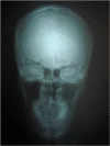
We present a 30-year-old South Indian man who presented with complaints of left sided headache and facial pain for past 3 months, severe for past 10 days. On physical examination, right side of the face appeared normal. Left side of the face showed signs of hemi atrophy with minimal drooping of left eyelid. All Systems were found to be normal. Routine blood and urine investigations results were within normal limits. X-ray chest revealed no abnormalities and x-ray skull showed both sides equal. Computerized tomogram of the brain showed left minimal sub dural hygroma with no midline shift, and no evidence of cerebral edema or cerebral atrophy. Nerve conduction study showed features suggestive of trigeminal neuralgia. MRI of the skull base was also normal and showed no evidence of trigeminal nerve compression. Interestingly, he had minimal response to analgesics, steroids, and propranolol, but showed immediate response to carbamazepine. Hence this patient indeed had Parry Romberg syndrome: Hemi facial atrophy with trigeminal neuralgia.
Figures


References
-
- Neville BW, Damm DD, Allen CN, Bouqout JE. Facilio Sugerica: Patologia oral e Maxilofacial. In: Guanabara Koogan., editor. 5. Vol. 1. Rio de Janeiro: Nike and Lidman; 1998. pp. 35–42.
Publication types
LinkOut - more resources
Full Text Sources
Miscellaneous

