

Intra-hepatic splenosis as an unexpected cause of a focal liver lesion in a patient with hepatitis C and liver cirrhosis: a case report
- PMID: 19830070
- PMCID: PMC2740154
- DOI: 10.4076/1757-1626-2-8335
Intra-hepatic splenosis as an unexpected cause of a focal liver lesion in a patient with hepatitis C and liver cirrhosis: a case report
Abstract
Introduction: Splenosis is the heterotopic autotransplantation of splenic tissue, mostly found after splenic trauma or surgery in the abdominal, pelvic or thoracic cavity. Here we report a patient with a history of splenectomy after polytrauma with chronic hepatitis C and liver cirrhosis presenting with an hepatic mass of unknown origin.
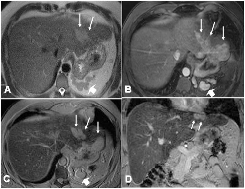
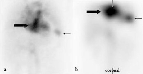
Case presentation: The lesion could not be exactly classified by ultrasound, computed tomography, angiography and biopsy, classical features of malignancy were not fulfilled, and on the other hand a neoplastic process could neither be excluded. After revision of a MRI performed in our centre it appeared that the liver mass contrasted in the same way as the remaining accessory spleens in the left upper quadrant. A selective Tc-99m-labelled heat-denatured autologous red blood cells scintigraphy of the spleen was performed and showed both the accessory spleens in the left upper quadrant and spleen-typical tissue in projection to the left liver lobe and confirmed the diagnosis of splenosis.
Conclusion: Although intrahepatic splenosis represents an extremely rare condition, this diagnosis should always be taken into consideration in patients with history of abdominal trauma with splenic involvement presenting with an indeterminate focal liver lesion. The diagnosis of splenosis may then be reliably confirmed by Tc-99m-DRBC scintigraphy.
Figures
Similar articles
-
Management of intrahepatic splenosis:a case report and review of the literature.World J Surg Oncol. 2018 Jun 28;16(1):119. doi: 10.1186/s12957-018-1419-1. World J Surg Oncol. 2018. PMID: 29954390 Free PMC article. Review.
-
Intrahepatic splenosis mimicking liver cancer: report of a case and review of literature.Int J Clin Exp Pathol. 2015 Jan 1;8(1):1031-5. eCollection 2015. Int J Clin Exp Pathol. 2015. PMID: 25755814 Free PMC article. Review.
-
Intrahepatic and intra-abdominal splenosis: A case report and review of literature.World J Hepatol. 2019 Dec 27;11(12):773-779. doi: 10.4254/wjh.v11.i12.773. World J Hepatol. 2019. PMID: 31966909 Free PMC article.
-
Intrahepatic Splenosis: A Rare Case.Curr Med Imaging. 2023;19(6):640-643. doi: 10.2174/1573405619666221212153639. Curr Med Imaging. 2023. PMID: 36515034
-
Hepatic splenosis presenting as arterialised liver lesion in a patient with NASH.Eur Rev Med Pharmacol Sci. 2013 Nov;17(21):2853-6. Eur Rev Med Pharmacol Sci. 2013. PMID: 24254551
Cited by
-
Solitary perihepatic splenosis mimicking liver lesion: a case report and literature review.Medicine (Baltimore). 2015 Mar;94(9):e586. doi: 10.1097/MD.0000000000000586. Medicine (Baltimore). 2015. PMID: 25738479 Free PMC article.
-
Primary splenic lymphoma on top of intrahepatic splenosis: A unique case report.Radiol Case Rep. 2022 Jun 13;17(8):2850-2854. doi: 10.1016/j.radcr.2022.02.064. eCollection 2022 Aug. Radiol Case Rep. 2022. PMID: 35782406 Free PMC article.
-
Management of intrahepatic splenosis:a case report and review of the literature.World J Surg Oncol. 2018 Jun 28;16(1):119. doi: 10.1186/s12957-018-1419-1. World J Surg Oncol. 2018. PMID: 29954390 Free PMC article. Review.
-
Ectopic Spleen Tissue - an Underestimated Differential Diagnosis of a Hypervascularised Liver Tumour.Viszeralmedizin. 2015 Dec;31(6):445-7. doi: 10.1159/000442115. Epub 2015 Dec 2. Viszeralmedizin. 2015. PMID: 26889148 Free PMC article.
-
Intrahepatic splenosis mimicking liver cancer: report of a case and review of literature.Int J Clin Exp Pathol. 2015 Jan 1;8(1):1031-5. eCollection 2015. Int J Clin Exp Pathol. 2015. PMID: 25755814 Free PMC article. Review.
References
-
- Gruen DR, Gollub MJ. Intrahepatic splenosis mimicking hepatic adenoma. AJR. 1997;168:725–726. - PubMed
-
- Lok AS, Seeff LB, Morgan TR, di Bisceglie AM, Sterling RK, Curto TM, Everson GT, Lindsay KL, Lee WM, Bonkovsky HL, Dienstag JL, Ghany MG, Morishima C, Goodman ZD, HALT-C Trial Group Incidence of hepatocellular carcinoma and associated risk factors in hepatitis C-related advanced liver disease. Gastroenterology. 2009;136:138–148. doi: 10.1053/j.gastro.2008.09.014. - DOI - PMC - PubMed
Publication types
LinkOut - more resources
Full Text Sources
