. 2009 Dec 15;108(6):1389-98.
doi: 10.1002/jcb.22371.

PDEF is a negative regulator of colon cancer cell growth and migration

Affiliations

PDEF is a negative regulator of colon cancer cell growth and migration

Omar Moussa et al. J Cell Biochem. .

Abstract

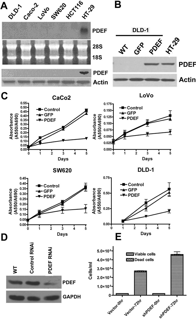
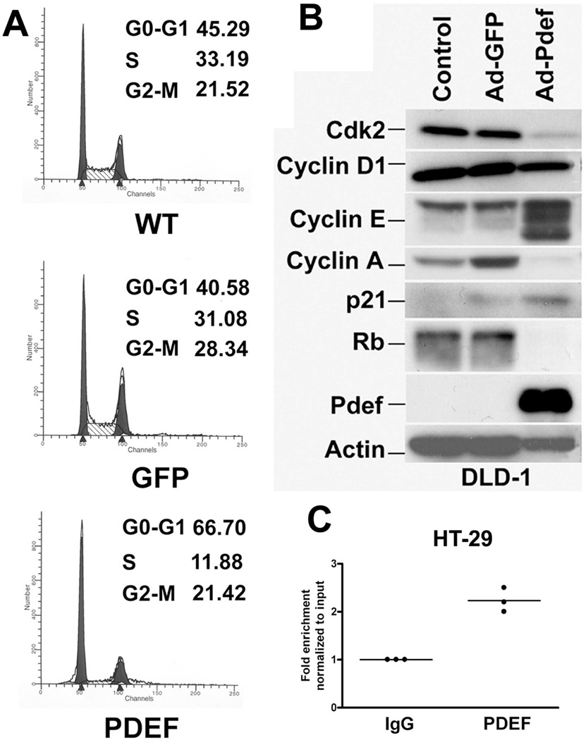
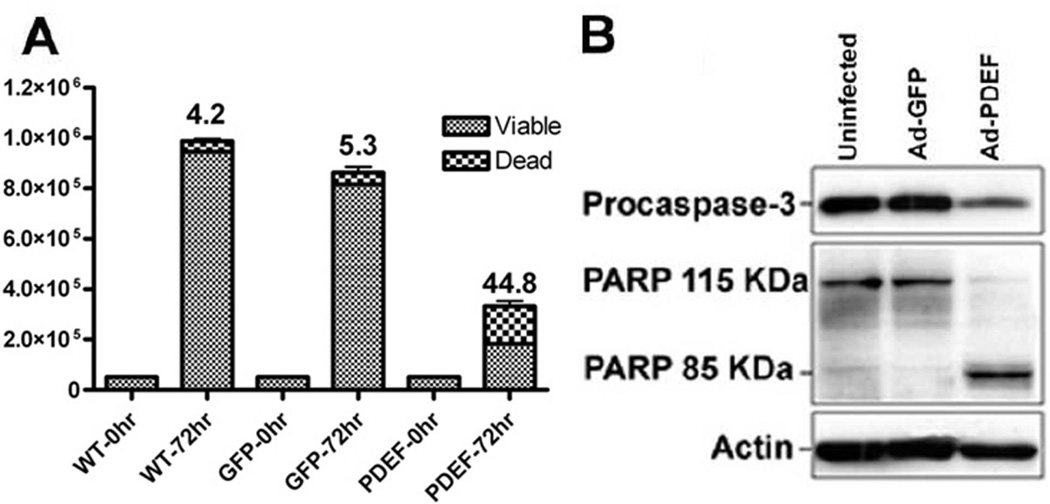
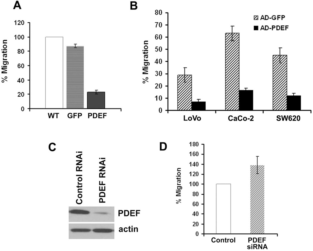
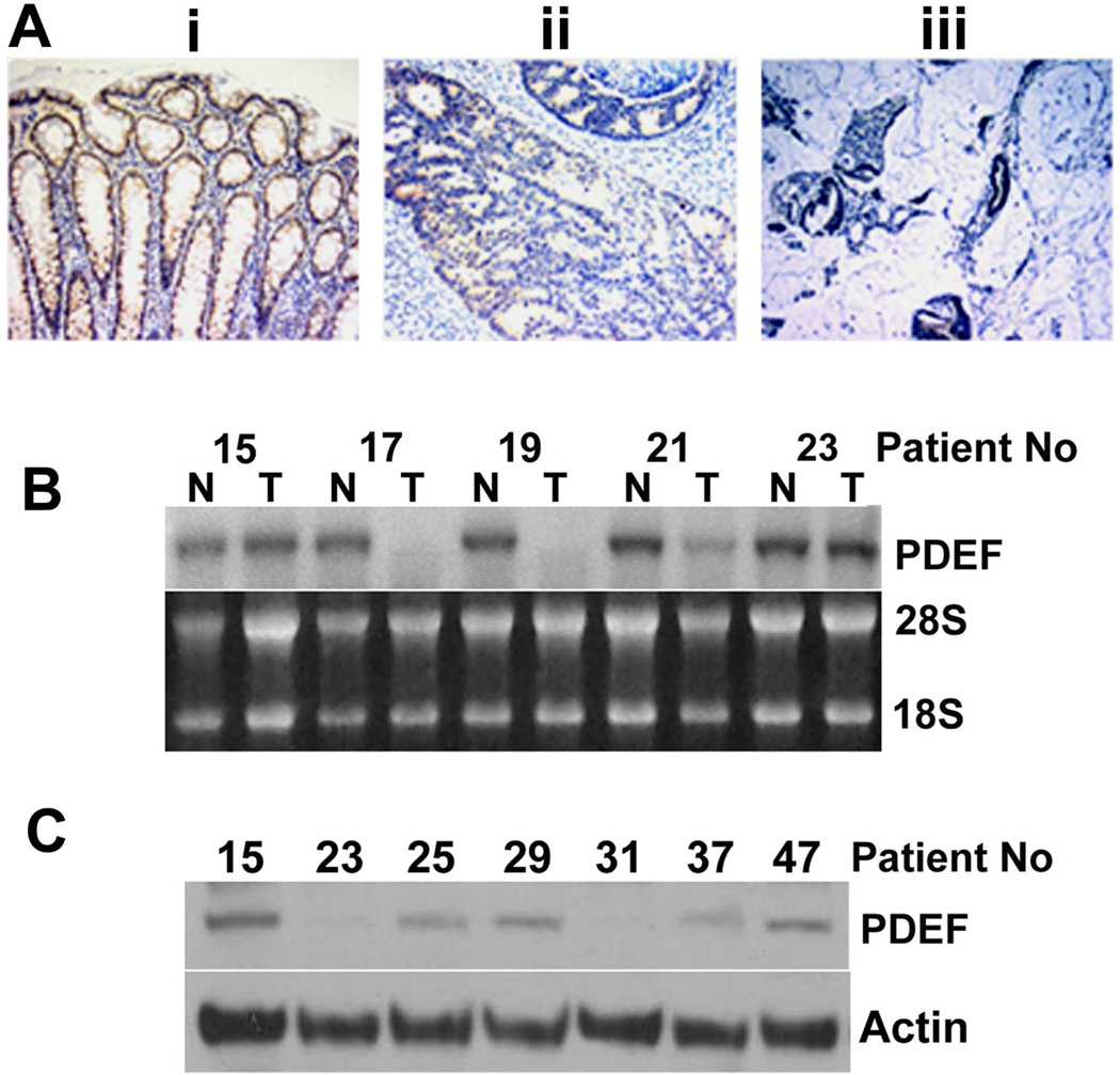
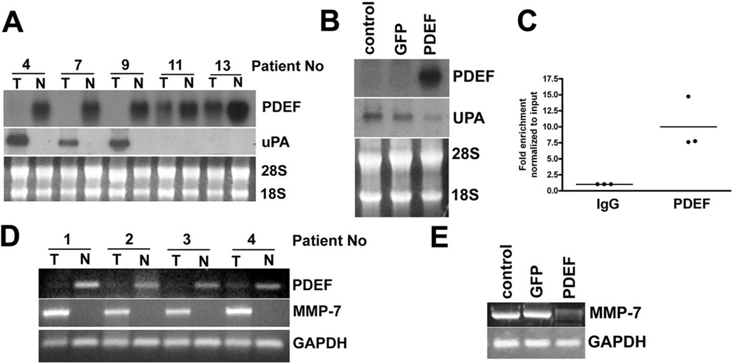
ETS is a family of transcriptional regulators with functions in most biological processes. Dysregulated ETS factor function leads to altered expression of multiple genes that play critical roles in many of the processes required for cancer progression. While the Ets family gene, prostate-derived ETS factor (PDEF), is expressed in epithelial tissues including prostate, breast, and colon, PDEF protein expression has been found to be reduced or lost during prostate and breast cancer progression. The goal of this study was to examine the expression and biologic impact of altered PDEF expression in colon cancer. PDEF mRNA and protein are not detectable in several colon-cancer-derived cell lines. Re-expression of PDEF in colon cancer cells inhibits growth and migration. Growth affects are due to altered cellular proliferation, indicated by increased altered cell population in G(1) and S phases of the cell cycle, as well as increased apoptosis. Relevant to its modulation of growth and migration phenotypes, PDEF expression resulted in altered expression of genes with established roles in cell cycle, motility, and invasion. Furthermore, chromatin immunoprecipitation studies show that p21 and urokinase plasminogen activator (uPA) are direct PDEF transcriptional targets. While non-tumor colon epithelium expresses PDEF mRNA and protein, the majority of tumors showed decreased mRNA and/or protein expression. In human tumor tissue samples, PDEF expression was inversely correlated with the expression levels of uPA. Collectively, the data support the model that PDEF is a negative regulator of tumor progression by modulating the expression of growth and migration promoting genes.


Figures

Figure 1
Figure 1. PDEF is lost in invasive colon cancer cell lines and re-expression of PDEF leads to growth changes in colon cancer cells
(A) Northern and Western blot analyses of RNA and protein prepared from the indicated colon cancer cell lines. Ethidium bromide staining of 28S and 18S rRNA or actin levels are provided as a controls. (B) Western blot analysis of PDEF expression level after infection of DLD-1 colon cells and endogenous expression level observed in HT-29 cells. Actin levels provided as a control. (C) CaCo-2, LoVo-2, SW680 and DLD-1 cells were untreated (control), or infected with either Ad-GFP or Ad-PDEF and cellular growth was monitored by MTT assay for 5 days. (D) Western blot analysis of PDEF protein expression in HT-29 colon cancer cells and in HT-29 expressing vector (RNAi control) and short hairpin RNA (shRNA) PDEF. Actin levels provided as a control. (E) Number of HT-29 colon cancer cells expressing vector alone or PDEF shRNA measured after 72 hours (p<0.05).
Figure 2
Figure 2. PDEF expression results in altered cell cycle progression and expression of cell cycle regulatory genes
(A) Cell cycle analysis of untreated DLD-1 cells or cells treated with Ad-GFP or Ad-PDEF and harvested 36 hours after infection. (B) Western blot analysis of total protein isolated from cells infected with Ad-GFP or Ad-PDEF using antibodies against the indicated cell cycle proteins. Blots were re-hybridized using antibodies against PDEF and β-Actin. Control lanes represent uninfected cells. (C) PDEF is bound to the p21 expression in vivo. Chromatin immuno-precipitation (CHIP) analysis using chromatin prepared from the HT-29 cell line. Q-PCR analysis using primers for the p21 promoter and chromatin immunoprecipitation with a PDEF-specific antibody, compared to IgG control antibody.
Figure 3
Figure 3. PDEF expression increases apoptosis
(A) Quantitation of trypan blue cell viability assays of parental (uninfected) DLD-1 cells, or cells infected with Ad-GFP or Ad-PDEF. The columns represent the average values for apoptotic index, the percentage of apoptotic vs. total cell number. Results are statistically significant with a p-value <0.05. (B) Western blot analysis of procaspase 3 and PARP expression levels in DLD-1 cells infected with Ad-GFP or Ad-PDEF. Actin levels provided as a control.
Figure 4
Figure 4. PDEF expression levels alter cell migration
The effect of PDEF expression on the migration of colon cancer cells was measured by transwell assay. (A) DLD-1 cells were untreated or infected with Ad-GFP or Ad-PDEF and migration measured by transwell assays. Data is representative of three experiments performed in triplicate. (B) LoVo, CaCo-2, and SW620 cells were infected with Ad-GFP or Ad-PDEF. Cell migration was measured by transwell migration assay. (C) Western blot analysis of PDEF protein expression in HT-29 colon cancer cells and in HT-29 expressing short hairpin RNA (shRNA) PDEF and control vectors. Actin levels provided as a control. (D) Cell migration of HT-29 colon cancer cells and of HT-29 cells expressing PDEF shRNA. Each of the measured differences were statistically significant with a p-value <0.05 by students t-test.
Figure 5
Figure 5. PDEF expression is reduced in colon cancer tissue
(A) Immunohistochemical analysis of PDEF expression in colon tissue: (i) non-tumor, (ii) tumor and (iii) invasive mucinous portion of tumor. Panels are from the same section. (Magnification, X400). Data representative of 5 of 6 cases. (B) Northern blot analysis for PDEF mRNA in a series of matched tumor (T) and non-tumor (N) tissues from the same patient (indicated by numbers). Data representative of 25 matched tumor/non-tumor samples. Ethidium bromide staining of 28S and 18S rRNA provided as control. (C) Western blot analysis of PDEF protein in 7 tumors that retain PDEF mRNA expression. Actin levels provided as a control.
Figure 6
Figure 6. PDEF loss alters the expression of the cancer associated genes, uPA and MMP-7
(A) Northern blot analysis of RNA prepared from tumor tissue and matched adjacent non-tumor tissues. 28S and 18S RNA provided as control for loading and RNA integrity. Data is representative of 25 tumor and non-tumor pairs examined. (B) Northern blot analysis of RNA prepared from parental DLD-1 cells and DLD-1 cells infected with Ad-GFP or Ad-PDEF. Ethidium bromide staining of 28S and 18S provided as a control. (C) Chromatin immuno-precipitation (CHIP) analysis using chromatin prepared from the HT-29 cell line. Q-PCR analysis using primers for the uPA promoter and chromatin immunoprecipitation with a PDEF-specific antibody, compared to IgG control antibody. (D) RT-PCR analysis MMP-7 mRNA in RNA prepared from tumor tissue and matched adjacent non-tumor tissues. GAPDH RT-PCR provided as a control. Data representative of 25 matched samples. (E) RT-PCR analyses of MMP-7 mRNA in RNA prepared from uninfected DLD-1 cells and cells infected with Ad-GFP or Ad-PDEF. GAPDH provided as control.

References

    1. Barker N, van de Wetering M, Clevers H. The intestinal stem cell. Genes Dev. 2008;22:1856–1864. - PMC - PubMed
    1. Beier F, Taylor AC, LuValle P. The Raf-1/MEK/ERK pathway regulates the expression of the p21(Cip1/Waf1) gene in chondrocytes. J Biol Chem. 1999;274:30273–30279. - PubMed
    1. Carbone GM, Napoli S, Valentini A, Cavalli F, Watson DK, Catapano CV. Triplex DNA-mediated downregulation of Ets2 expression results in growth inhibition and apoptosis in human prostate cancer cells. Nucleic Acids Res. 2004;32:4358–4367. - PMC - PubMed
    1. Cheng K, Xie G, Raufman JP. Matrix metalloproteinase-7-catalyzed release of HB-EGF mediates deoxycholyltaurine-induced proliferation of a human colon cancer cell line. Biochem Pharmacol. 2007;73:1001–1012. - PMC - PubMed
    1. Cohen GM. Caspases: the executioners of apoptosis. Biochem J. 1997;326:1–16. - PMC - PubMed

Publication types

MeSH terms

LinkOut - more resources