. 2010 Jan;40(1):36-46.
doi: 10.1002/eji.200939748.

HIV gag protein is efficiently cross-presented when targeted with an antibody towards the DEC-205 receptor in Flt3 ligand-mobilized murine DC

Affiliations

HIV gag protein is efficiently cross-presented when targeted with an antibody towards the DEC-205 receptor in Flt3 ligand-mobilized murine DC

Leonia Bozzacco et al. Eur J Immunol. 2010 Jan.

Abstract

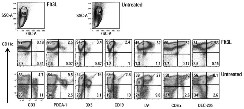
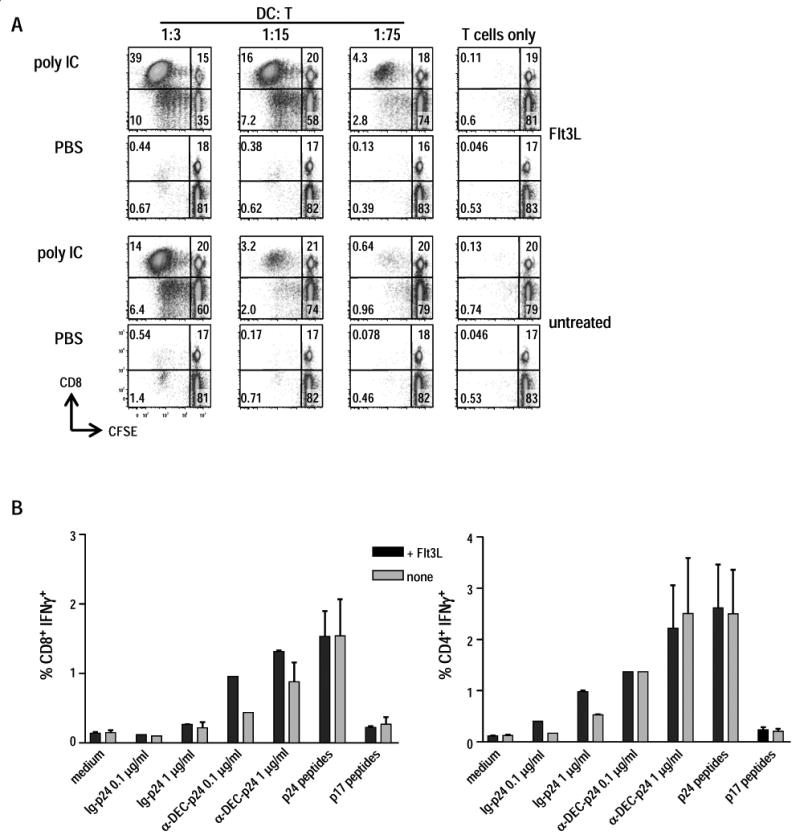
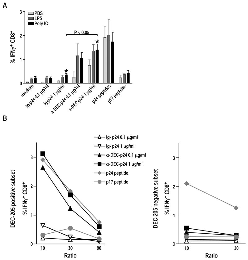
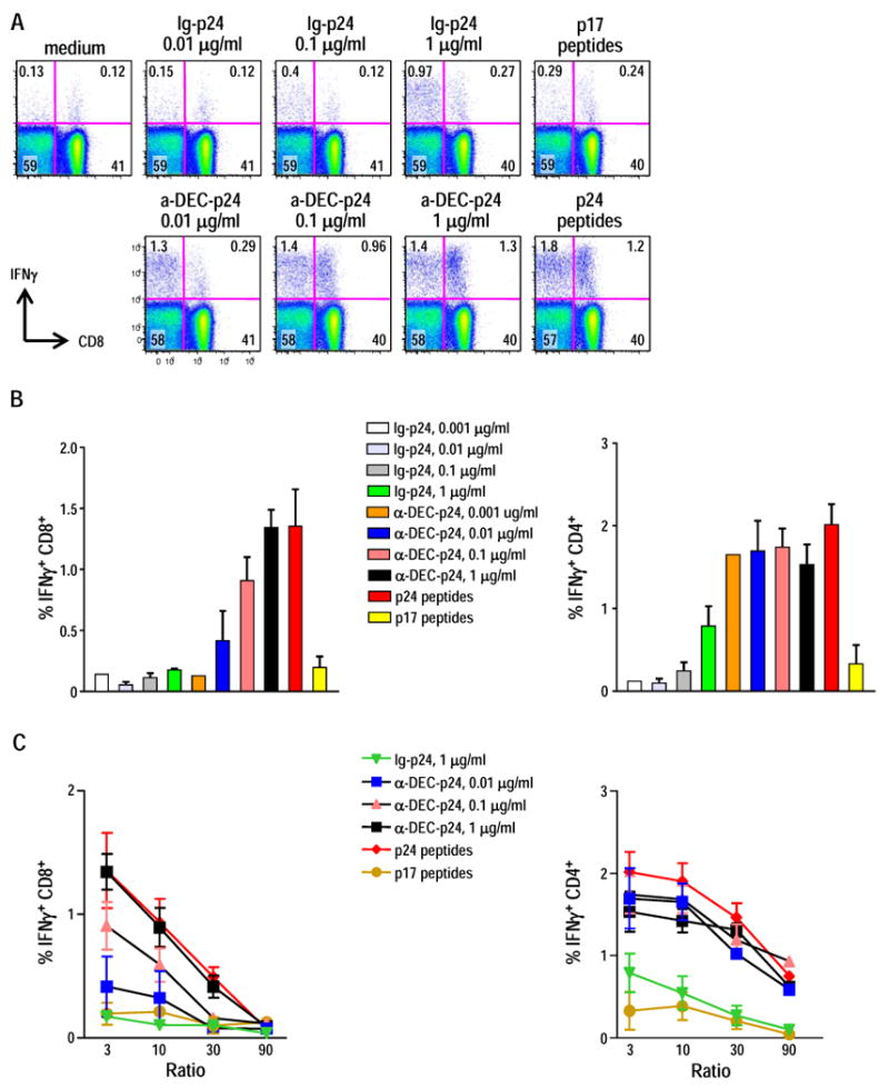
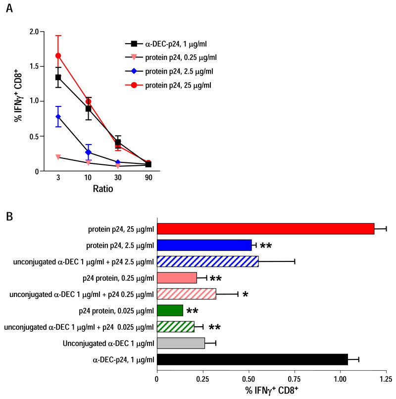
DC present exogenous proteins to MHC class I-restricted CD8+ T cells. This function does not require endogenous antigen synthesis within DC, providing the potential to elicit CD8+ T-cell responses to immune complexes, inactivated microbes, dying cells, and proteins such as OVA. In mice, the CD8+ or DEC-205+ DC are specialized for cross-presentation, and this subset can be increased 10-fold in numbers following Fms-like tyrosine kinase 3 ligand (Flt3L) treatment in vivo. Therefore, we studied cross-presentation by abundant Flt3L DC using HIV gag protein. When enriched by positive selection with anti-CD11c beads, cells from Flt3L mice are not only more abundant but are also more highly enriched in CD11chigh DC, particularly the DEC-205+ subset. DC cross-present HIV gag to primed CD8+ T cells, but when the antigen is delivered within an antibody to DEC-205 receptor, cross-presentation becomes 100-fold more efficient than non-targeted antigen. This finding requires gag to be engineered into anti-DEC antibody, not just mixed with antibody. Flt3L DC are a valuable tool to study cross-presentation, since their use overcomes the obstacle posed by the low number of cross-presenting DC in the steady state. These findings support future experiments to use Flt3L to enhance presentation of DC-targeted vaccines.


Conflict of interest statement

Conflict of interest. RMS is a paid scientific consultant to Celldex Therapeutics which is developing DEC-205-targeted vaccines.

Figures

Figure 1
Figure 1. Cell surface markers of splenic CD11c+ DC by flow cytometry
Splenic CD11c+ cells were enriched by positive selection on anti-CD11c beads from CxB6 F1 mice that had been injected with B16 Flt3L melanoma cells or from untreated F1 mice. A six-color flow cytometry panel was used to simultaneously analyze surface markers on CD11c-selected cells. Appropriate isotype-matched, non-binding control Ig's were used to set the quadrant lines (not shown). DC were analyzed by gating against low scatter debris, followed by a live/dead exclusion gate. Analysis is representative of three independent experiments that all examined pooled splenic DC from at least two mice.
Figure 2
Figure 2. Comparision of antigen-presentation by Flt3L and untreated CD11c+ DC
(A) B16 Flt3L-injected or untreated CxB6 F1 mice were stimulated with 50 μg of poly IC for 15 h and allo-stimulatory capacity of splenic CD11c+ DC was compared. Both populations of DC were fixed with 1% para-formaldehyde, washed 3 times, and cultured at various numbers in the presence of a constant number (3×10⁵) of T cells enriched from allogenic C57BL/6 mice. T cell proliferation was detected after 4 days of culture by CFSE dilution of CD8+ and CD4+ T cells. Similar results were obtained using allogeneic T cells from the SJL mouse strain. Data are representative of two independent experiments. (B) Splenic CD11c+ cells purified from Flt3L or untreated F1 mice were pulsed in vitro for 4-5 h with anti-DEC-p24, control Ig-p24 at 0.1 and 1 μg/ml, p24 or p17 15-mer peptides mix (1 μg/ml), followed by 25 μg/ml of poly IC. Fifteen hours later, 1×10⁶ washed DC were added to 3×10⁶ T cells for 6 h in the presence of BFA (10 μg/ml) to detect IFN-γ secretion by intracellular cytokine staining. The T cells were isolated from F1 mice that were primed with Adenovirus gag and boosted with anti-DEC-p24 and poly IC (Methods). Data show mean ± SD of the percentage of CD8+ IFN-γ+ T cells (left) or CD4+ IFN-γ+ T cells (right) representative of two independent experiments.
Figure 3
Figure 3. Cross-presentation of anti-DEC-gag p24 is enhanced by poly IC and is restricted to DEC-205+ DC
(A) Splenic Flt3L-mobilized CD11c+ DC were pulsed in vitro with 0.1 and 1 μg/ml of anti-DEC-p24, control Ig-p24, or p24 or p17 15-mer peptides (1 μg/ml). Five hours later, medium or LPS (0.1 μg/ml) or poly IC (25 μg/ml) were added. After overnight culture, the yields of cells were comparable. The cells were washed and added for 6 h to HIV gag primed T cells at a ratio of 1:3 with 10 μg/ml BFA. Data show mean ± SD percentage of CD8+ IFN-γ+ T cells pooled from three independent experiments. *p<0.05, paired Student's t-test, 1 μg/ml of anti-DEC-p24 vs. 1 μg/ml of control-Ig-p24 after maturation with poly IC. (B) CD11c+ cells from mice injected with B16 Flt3L melanoma cells were enriched by MACS positive selection and then separated into DEC-205+ and DEC-205- CD11chigh DC subsets by FACS. Sorted cells were washed and pulsed with either medium, anti-DEC-p24 vs. control Ig-p24 at 0.1 or 1 μg/ml, or p24 vs p17 peptides at 1 μg/ml. Five hours later, without washing off the antigens, poly IC at 25 μg/ml was added. After 15 h, graded doses of DEC-205+ and DEC205- cells (x-axis for DC:T cell ratio) were added to 3×10⁶ HIV gag primed T cells, and IFN-γ secretion was assessed by intracellular cytokine staining 6 h later. Frequencies of CD8+ IFN-γ+ T cells from two independent experiments are shown after stimulation by DEC-205+ (left panel) and DEC-205- cells (right panel).
Figure 4
Figure 4. Efficient gag-presentation to CD8+ and CD4+ T cells following in vitro targeting of Flt3L DC with anti-DEC-gag p24 mAb
(A) Splenic Flt3L CD11c+ DC pulsed with anti-DEC-p24, control Ig-p24, p24 or p17 peptides at the concentration indicated, matured overnight with poly IC 25 μg/ml were used to stimulate IFN-γ secretion by HIV gag primed T cell in a 6 h assay at 1:3 DC: T cell ratio. Dot plots are representative of three independent experiments. (B) As in A, but Flt3L DC were pulsed with limiting dilutions of anti-DEC-p24 and control Ig-p24, matured with poly IC and cocultured for 6 h with primed HIV gag T cells. IFN-γ secretion from CD8+ T cells and CD4+ T cells was assessed by intracellular cytokine staining. Data show mean ± SD for the percentage of CD8+ IFN-γ+ T cells (left) or CD4+ IFN-γ+ T cells (right) from four independent experiments. (C) Graded doses of Flt3L DC, pulsed and matured as described in B, were cocultured for 6 h with primed HIV gag T cells (x-axis for DC:T cell ratio). IFN-γ secretion from CD8+ T cells (left) and CD4+ T cells (right) was assessed by intracellular cytokines. Mean ± SD of the frequencies of IFN-γ producing CD8+ and CD4+ T cells in three to four independent experiments are indicated.
Figure 5
Figure 5. Conjugation of DEC-205 to HIV gag, but not DEC-205 itself, greatly enhances cross-presentation of soluble HIV gag p24 protein
(A) Splenic Flt3L CD11c+ DC were pulsed with 1 μg/ml of anti-DEC-p24 or increasing doses of HIV gag p24 soluble protein. The DC were matured with 25 μg/ml poly IC and added at various doses to stimulate primed HIV gag T cells in a 6 h coculture (x-axis for DC:T cell ratio). IFN-γ secretion from CD8+ T cells was assessed by intracellular cytokine staining. Data show mean ± SD of three independent experiments. (B) Splenic Flt3L CD11c+ DC were pulsed with 1 μg/ml of anti-DEC-p24, or 1 μg/ml of unconjugated anti-DEC, or different combinations of HIV gag p24 protein and unconjugated anti-DEC, or limiting doses of HIV gag p24 protein. As above, the DC were matured with poly IC and added at various doses to stimulate primed HIV gag T cells in a 6 h coculture (x-axis for DC:T cell ratio). IFN-γ secretion from CD8+ T cells was assessed by intracellular cytokine staining. Data show mean ± SD of two independent experiments. *p<0.05, **p<0.01, paired Student's t-test; 1 ug/ml of anti-DEC-p24 vs. other samples.

References

    1. Heath WR, Belz GT, Behrens GM, Smith CM, Forehan SP, Parish IA, Davey GM, Wilson NS, Carbone FR, Villadangos JA. Cross-presentation, dendritic cell subsets, and the generation of immunity to cellular antigens. Immunol Rev. 2004;199:9–26. - PubMed
    1. Regnault A, Lankar D, Lacabanne V, Rodriguez A, Thery C, Rescigno M, Saito T, Verbeek S, Bonnerot C, Ricciardi-Castagnoli P, Amigorena S. Fcγ receptor-mediated induction of dendritic cell maturation and major histocompatibility complex class I-restricted antigen presentation after immune complex internalization. J Exp Med. 1999;189:371–380. - PMC - PubMed
    1. Kalergis AM, Ravetch JV. Inducing tumor immunity through the selective engagement of activating Fcγ receptors on dendritic cells. J Exp Med. 2002;195:1653–1659. - PMC - PubMed
    1. Den Haan JM, Bevan MJ. Constitutive versus activation-dependent cross-presentation of immune complexes by CD8(+) and CD8(-) dendritic cells in vivo. J Exp Med. 2002;196:817–827. - PMC - PubMed
    1. Liu K, Iyoda T, Saternus M, Kimura K, Inaba K, Steinman RM. Immune tolerance after delivery of dying cells to dendritic cells in situ. J Exp Med. 2002;196:1091–1097. - PMC - PubMed

Publication types

LinkOut - more resources