. 2009 Dec;47(12):805-14.
doi: 10.1002/dvg.20564.

Generation of aggrecan-CreERT2 knockin mice for inducible Cre activity in adult cartilage

Affiliations

Generation of aggrecan-CreERT2 knockin mice for inducible Cre activity in adult cartilage

Stephen P Henry et al. Genesis. 2009 Dec.

Abstract

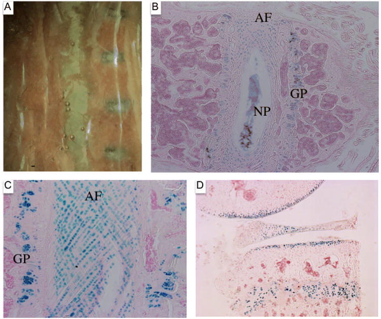
The function of cartilage in the adult is dependent on a host of regulatory molecules such as growth factors, extracellular matrix, enzymes, signaling molecules, and transcription factors. However, germline mutations in some genes that are expressed in adult cartilage lead to embryonic or perinatal lethality. To examine the function of these and other genes postnatally, we have generated a targeted mouse by homologous recombination that "knocks in" the inducible Cre recombinase construct, CreERT2, in the 3' untranslated region of the endogenous mouse aggrecan gene (Agc1(tm(IRES-creERT2))). The properties and efficiency of the inducible cre recombinase were tested by examining X-gal staining of tissues from embryos as well as growing and adult Agc1(tm(IRES-creERT2)/+);Rosa 26R mice. These mice were injected with the inducer, tamoxifen, at different time points during embryonic development and postnatally up to 6 months of age. Strong X-gal staining was observed in growth plate and articular cartilage as well as the fibrocartilage of meniscus, trachea, and intervertebral discs reproducing the pattern of endogenous aggrecan gene expression. In conclusion, we have generated a mouse model in which genes implicated in cartilage degenerative diseases can be inactivated in a spatial and temporal fashion in postnatal and adult mice.


Figures

Figure 1
Figure 1
Gene-targeting strategy for the Aggrecan-CreERT2 allele. Top panel displays the genomic structure of the mouse aggrecan gene. Middle panel shows the targeting vector. Bottom Panel depicts the targeted allele whereby IRES-CreERT2 and an FRT-flanked Pgk-Neo cassette (in reverse transcriptional orientation to aggrecan) is recombined into the 3′ untranslated region (3′ UTR) of the aggrecan gene. EcoRV restriction enzyme digestion is used in Southern blotting analysis to identify correctly targeted ES cell cones. Both the 5′ and 3′ probes (position shown in the middle panel) detect a 13.5 kb fragment with the wild-type allele. The targeted allele generates a 5.4 or 9.6 kb fragment detected by the 5′ or 3′ probe, respectively. At the bottom of the panel a targeted ES clone (3H11) is shown to the left in a Southern blot hybridized with the radiolabeled 5′ probe. The Southern blot of the 3′ probe with the same clone is shown to the right. RV: EcoRV sites. TK: MC1-TK. Neo: Pgk-Neo. FRT: FRT sites.
Figure 2
Figure 2
Pregnant mothers were injected intraperitoneally once with 4OH-tamoxifen (total of 3mgs injected per mother) or vehicle control (oil). Females were sacrificed 24 hours later, and the embryos were collected and stained with X-gal. All embryos in the figure are genotype Agc1tm(IRES-CreERT2/+); R26R. A: No X-gal positive signal was detected at embryonic day 12.5 (E12.5), but X-gal positive signal was detected in the forelimb at E13.0 (arrow). At E13.5, signal was detected in the cartilage of the axial and appendicular skeleton. B: Left, Agc1tm(IRES-CreERT2/+); R26R E15.5 embryo harvested from a vehicle control (oil) injected mother and stained with X-gal; right, E15.5 embryo of the same genotype obtained from a different mother injected with 4OH-tamoxifen and stained with X-gal. Positive X-gal staining is detected in cartilage only in the embryo harvested from the 4OH-tamoxifen injected female. C: Dame injected one day prior to delivery, and pups were sacrificed at postnatal day 2 (P2). A P2 Agc1tm(IRES-CreERT2/+); R26R pup whose skin had been removed, was stained with X-gal showing positive signal in cartilage structures. D: Histological section of an X-gal stained ulna and radius growth plate from an E15.5 Agc1tm(IRES-CreERT2/+); R26R embryo harvested from a tamoxifen injected mother. Positive X-gal staining is detected in both the proliferating and hypertrophic chondrocytes. E: Histological section of an ulna from an E15.5 Agc1tm(IRES-CreERT2/+); R26R embryo harvested from an oil injected mother that was subjected to RNA in situ hybridization with a digoxigenin labeled aggrecan mRNA probe. Aggrecan mRNA was in detected in the proliferating chondrocytes of the ulna growth plate but was not detected in the hypertrophic chondrocytes.
Figure 3
Figure 3
Tissues harvested from 6-month-old Agc1tm(IRES-CreERT2/+); R26R mice that were injected intraperitoneally with either tamoxifen (1.5 mgs tamoxifen/10 gms of mouse weight) or vehicle alone (oil), starting at 6 months for 3 consecutive days and sacrificed 3 days later. All tissues were mildly fixed and bathed in X-gal solution. A: Lateral view of the knee joints from two animals injected with tamoxifen (bottom two joints of panel) showing X-gal staining in the growth plate (*) and articular (arrow to tibial plateau) cartilage, but no positive X-gal staining is detected in the knee joint from the animal injected with oil (top of panel). B: Top view of tibia and femur from panel A showing the mouse tissue from oil injected mouse to the left, and tamoxifen injected mouse to the right. Positive X-gal staining is detected in the growth plate (*), articular cartilage of femoral condyle (arrow head), and tibial plateau (arrow), as well as the meniscus (“m”) from the tamoxifen-injected mouse. C: Lateral view of two ankles with skin and muscle removed (left, oil injected, -; right, tamoxifen, +). The positive X-gal staining at the tendon-bone junction is labeled with arrow. D: Dissected and cut trachea from a tamoxifen injected animal. E: Top view of rib cage from tamoxifen injected mouse bathed in X-gal (rib cartilage, arrow; trachea, arrow head; spine, asterisk). F: Cranial bones covering the skulls of either oil injected (left) or tamoxifen injected (right) animals were removed to expose the brain to X-gal solution. No X-gal staining was detected at the surface of the brain from the tamoxifen-injected animal, even though the nasal cartilage from the tamoxifen-injected animal was X-gal positive (right, arrow, +).
Figure 4
Figure 4
Histological sections of the knee joint from Agc1tm(IRES-CreERT2/+); R26R mice mouse injected with either tamoxifen or vehicle alone (oil) and stained with X-gal. A: Histological section of the knee joint from a 2.5 week old Agc1tm(IRES-CreERT2); Rosa26 mouse injected once with oil at 2 weeks and stained with X-gal. B: View of the articular cartilage in Panel A. No positive X-gal staining is observed in the growth plate or the articular cartilage. C: RNA in situ hybridization of a histological section with an aggrecan labeled digoxigenin probe showing the growth plate of an uninjected 2.5 Agc1tm(IRES-creERT2/+); R26R mouse. Aggrecan mRNA is detected in the reserve zone (RZ), the proliferative zone (PZ) and the upper hypertrophic zone (HZ). However, aggrecan mRNA is not detected in a large number of hypertrophic chondrocytes in the lower hypertrophic zone (arrows). D: Histological section of the knee joint from a 2.5 week old Agc1tm(IRES-CreERT2); R26R mouse injected once with tamoxifen at 2 weeks and stained with X-gal. Positive X-gal staining is observed in the resting, proliferative, and hypertrophic zones of the growth plate. E: View of panel D showing positive X-gal staining in all of the chondrocytes of the articular cartilage including the deep layers. F: Histological section of the knee joint from a 4.5 week old Agc1tm(IRES-CreERT2); R26R mouse injected once with tamoxifen at 4 weeks and stained with X-gal. G: View of Panel F focusing on the growth plate showing positive X-gal staining for over 90% of growth plate chondrocytes including all hypertrophic chondrocytes. H: View of Panel F focusing on the articular cartilage showing high number of X-gal positive chondrocytes in the uncalcified cartilage above the tidemark, but very little X-gal positive chondrocytes in the calcified cartilage below the tidemark (labeled as asterisk). Scale bar is 200 microns.
Figure 5
Figure 5
Histological sections of 6 month-old Agc1tm(IRES-creERT2/+); R26R knee joints. A: Histological section of the knee joint from a 6-month-old mouse hybridized with an antisense aggrecan digoxigenin labeled RNA probe. The aggrecan mRNA is detected in the growth plate cartilage (GP). In the articular cartilage (AC) overlying the secondary ossification center (SO), the aggrecan mRNA is detected in the chondrocytes of the uncalcified cartilage above the tidemark (see arrow), but not in the chondrocytes of the calcified cartilage below the tidemark. In the meniscus (ME), the aggrecan mRNA is detected only in the outer superficial chondrocytes of the meniscus (white arrowhead). B: The growth plate from a histological section of a 6 month-old Agc1tm(IRES-CreERT2); R26R mouse injected with tamoxifen and stained with X-gal and counterstained with nuclear fast red. Positive X-gal staining is detected in the chondrocytes of the growth plate. C: Histological section of the knee joint from a tamoxifen-injected 6-month-old Agc1tm(IRES-CreERT2); R26R stained with X-gal. Positive X-gal stained chondrocytes are observed in the uncalcified cartilage above the tidemark (see arrow), but not in the chondrocytes of the calcified cartilage below the tidemark. In the meniscus (ME), positive X-gal stained chondrocytes are only detected in the outer superficial chondrocytes of the meniscus (arrowhead). D: Histological section of the knee joint showing both the tibial and femoral articular cartilages from a 6 month-old Agc1tm(IRES-creERT2/+); R26R mouse injected with tamoxifen and stained with X-gal. X-gal positive chondrocytes are seen in the uncalcified cartilage above the tidemark (shown as curved black line), but not detected in the calcified cartilage (shown as asterisk). E: Histological section of the knee joint stained with X-gal from a 6 month-old Agc1tm(IRES-creERT2/+); R26R control mouse injected with vehicle alone (oil). No X-gal staining was observed in the cartilage.
Figure 6
Figure 6
The vertebral columns from tamoxifen and oil injected 6-month-old Agc1tm(IRES-creERT2/+); R26R mice were isolated and stained with X-gal. A: Vertebral column from oil injected mouse to the left and from tamoxifen injected animal to the right. B: Histological section of the vertebral column from the tamoxifen-injected 6-month-old Agc1tm(IRES-CreERT2); R26R mouse stained with X-gal and counterstained with nuclear fast red. Positive X-gal staining is detected in the growth plate (GP) of the vertebral body, the nucleus pulposus (NP), and the rings of the annulus fibrosis (AF). C: Positive X-gal staining of cells contained in the rings of the annulus fibrosis (AF) and the chondrocytes of the growth plate (GP) in the vertebral body. D: Histological section of the knee joint from a 1-year-old Agc1tm(IRES-CreERT2); R26R mouse injected with tamoxifen starting at one year and sacrificed one week later. The X-gal stained joint showed positive signal in the growth plate, articular cartilage, and the meniscus.

References

    1. Bruckner G, Grosche J, Hartlage-Rubsamen M, Schmidt S, Schachner M. Region and lamina-specific distribution of extracellular matrix proteoglycans, hyaluronan and tenascin-R in the mouse hippocampal formation. J Chem Neuroanat. 2003;26:37–50. - PubMed
    1. Chambers MG, Kuffner T, Cowan SK, Cheah KS, Mason RM. Expression of collagen and aggrecan genes in normal and osteoarthritic murine knee joints. Osteoarthritis Cartilage. 2002;10:51–61. - PubMed
    1. Chen M, Lichtler AC, Sheu TJ, Xie C, Zhang X, O'Keefe RJ, Chen D. Generation of a transgenic mouse model with chondrocyte-specific and tamoxifen-inducible expression of Cre recombinase. Genesis. 2007;45:44–50. - PMC - PubMed
    1. Costa C, Tortosa R, Domenech A, Vidal E, Pumarola M, Bassols A. Mapping of aggrecan, hyaluronic acid, heparan sulphate proteoglycans and aquaporin 4 in the central nervous system of the mouse. J Chem Neuroanat. 2007;33:111–123. - PubMed
    1. George SH, Gertsenstein M, Vintersten K, Korets-Smith E, Murphy J, Stevens ME, Haigh JJ, Nagy A. Developmental and adult phenotyping directly from mutant embryonic stem cells. Proc Natl Acad Sci U S A. 2007;104:4455–4460. - PMC - PubMed

Publication types

MeSH terms

LinkOut - more resources