. 2009 Nov;47(11):775-81.
doi: 10.1002/dvg.20562.

Generation and analysis of a mouse line harboring GFP in the Eomes/Tbr2 locus

Affiliations

Generation and analysis of a mouse line harboring GFP in the Eomes/Tbr2 locus

Sebastian J Arnold et al. Genesis. 2009 Nov.

Abstract

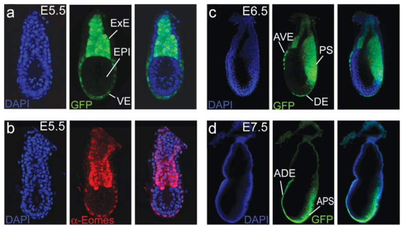
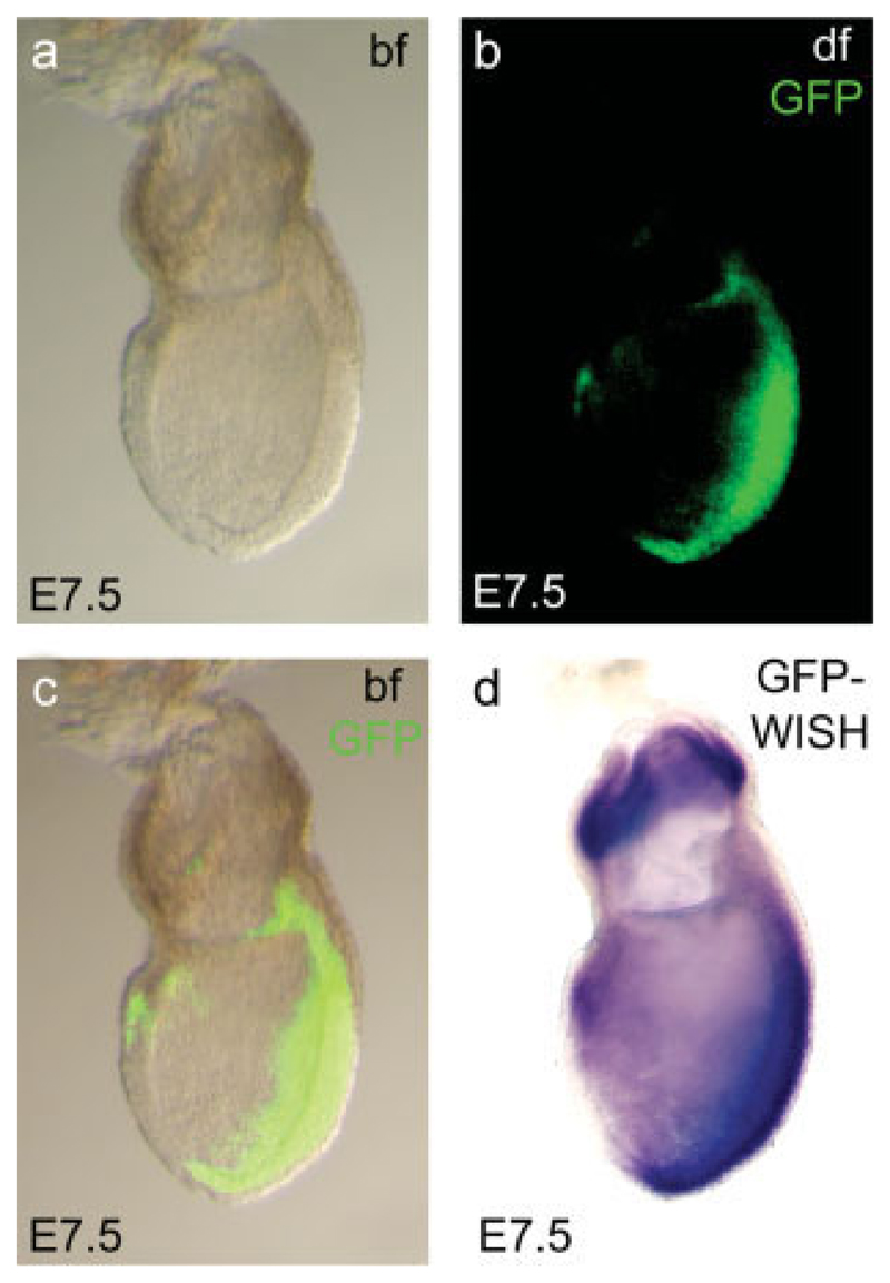
During mouse embryonic development, the T-box transcription factor Eomes/Tbr2 is expressed in highly dynamic patterns in various progenitor cell types. Those include the undifferentiated cells of the trophectoderm, ingressing nascent mesoderm at the primitive streak, and intermediate progenitor cells of the developing cerebral cortex. We generated an Eomes(GFP)- targeted allele to follow the highly dynamic patterns of Eomes expression and to allow for the identification of novel expression domains. We show that our novel allele recapitulates endogenous gene expression at known sites of expression and confirm our results by anti-Eomes immunofluorescent staining. Using this novel allele we were able to identify previously undocumented domains of Eomes expression within the visceral endoderm and at various locations in the developing and adult mouse brain.


Figures

Fig. 1
Fig. 1
Generation of the EomesGFP reporter allele. (a) Strategy to introduce EGFP into the first coding Exon 1 of the Eomes locus by homologous recombination in ES cells. A EGFP.pA cassette was placed into the transcriptional start site of the endogenous Eomes locus, followed by a removable, LoxP-flanked (blue arrows) selection cassette (PGK.neo). (b) Drug-resistant ES cell clones were screened by Southern Blot on EcoRV-digested DNA using a 3 external probe (indicated as red bar in a). (c) Correctly targeted clones were subjected to transient expression of Cre. Individual clones were analyzed by Southern blot to distinguish wildtype allele (wt, 15 kb), targeted allele (T, 7.0 kb), and reporter allele (R, 5.5 kb). (d) PCR genotyping distinguishes wildtype (wt, 302 bp) and EomesGFP reporter allele (R, 373 bp). (e) Genotyping of embryos from EomesGFP/+ intercrosses demonstrates lethality of EomeGFP/GFP embryos prior to E7.5. (f) EomesGFP/CA; Sox2.Cre epiblast-deleted embryos fail to form the mesoderm cell layer and are blocked in gastrulation as shown by cross-sections of E7.5 embryos. E, EagI; H, HindIII; S, SphI; RV, EcoRV; TK, thymidine kinase (negative selection marker).
Fig. 2
Fig. 2
EomesGFP expression in the early embryo. (a) At E5.5 EomesGFP expression is found in the extraembryonic ectoderm (ExE) and at reduced levels in the embryonic visceral endoderm (VE). The epiblast (EPI) is devoid of any GFP expression. (b) Immunofluorescence staining with an anti-Eomes antibody at E5.5 confirms the strong expression of endogenous Eomes protein within the ExE and at slightly reduced levels in the VE. (c) After onset of gastrulation at E6.5, EomesGFP is detected in the primitive streak (PS). Additional expression is found in remaining cells of the anterior visceral endoderm (AVE) and in nascent definitive endoderm (DE). (d) During later stages of gastrulation, at E7.5, GFP remains predominantly expressed in the anterior part of the PS (APS) and in cells of the anterior definitive endoderm (ADE). No or only very weak expression is found in the derivatives of the ExE, such as chorion and the ectoplacental cone.
Fig. 3
Fig. 3
Comparison of EomesGFP fluorescence and mRNA expression at late gastrulation. GFP fluorescence can be identified throughout the primitive streak region and in a few cells in the anterior endoderm of the E7.5 late gastrulation stage embryos (a–c). No GFP fluorescence can be detected within the extraembryonic ectoderm (b, c), despite highly abundant mRNA expression as demonstrated by whole mount in situ hybridization (WISH, d). bf, brightfield; df, darkfield.
Fig. 4
Fig. 4
EomesGFP expression during neurogenesis of the developing cerebral cortex. Coronal sections through the cortical fore-brain regions of E12 (a–d’) and E15 (e–h’) EomesGFP mice demonstrate gross overlap of endogenous Eomes protein expression and GFP. (b, b’) At E12, Eomes protein is found in cells of the pre-plate (arrow) and in few prolifera-tive cells in proximity to the ventricular surface (asterisk). (c) GFP expression extends more lateral (arrows in c’ and d’) than immuno-fluorescent staining. (f, f’) In the E15 cortex, Eomes protein is localized to the nuclei of the basal ventricular zone (VZ) and the sub-ventricular zone (SVZ). (g’, h’) Eomes protein expression is recapitulated by cytoplasmic GFP expression that extends marginally more toward the intermediate zone.
Fig. 5
Fig. 5
EomesGFP expression in the adult brain. Comparisons of Eomes immunofluorescent staining and EomesGFP at different locations in the adult (postnatal day 50, P50) mouse brain. (a) In the adult olfactory bulb, Eomes nuclear staining and cytoplasmic GFP expression are found in the mitral cell layer (arrow) and within the granular layer (asterisk). (b) Few Eomes and GFP-positive cells are located in the subepen-dymal zone, adjacent to the ventricles (asterisk) and (c) in the region of the ventral septum. (d) Weak expression of both Eomes protein and GFP expression is found in the internal granular cell layer of the adult cerebellum. (e) In the adult dentate gyrus, Eomes and GFP expression is seen in the subgranular zone, the site of proliferating progenitor cells.

References

    1. Arnold SJ, Hofmann UK, Bikoff EK, Robertson EJ. Pivotal roles for eomesodermin during axis formation, epithelium-to-mesen-chyme transition and endoderm specification in the mouse. Development. 2008a;135:501–511. - PMC - PubMed
    1. Arnold SJ, Huang GJ, Cheung AF, Era T, Nishikawa S, Bikoff EK, Molnar Z, Robertson EJ, Groszer M. The T-box transcription factor Eomes/Tbr2 regulates neurogenesis in the cortical subventricular zone. Genes Dev. 2008b;22:2479–2484. - PMC - PubMed
    1. Arnold SJ, Robertson EJ. Making a commitment: Cell lineage allocation and axis patterning in the early mouse embryo. Nat Rev Mol Cell Biol. 2009;10:91–103. - PubMed
    1. Bulfone A, Martinez S, Marigo V, Campanella M, Basile A, Quaderi N, Gattuso C, Rubenstein JL, Ballabio A. Expression pattern of the Tbr2 (Eomesodermin) gene during mouse and chick brain development. Mech Dev. 1999;84:133–138. - PubMed
    1. Chazaud C, Rossant J. Disruption of early proximodistal patterning and AVE formation in Apc mutants. Development. 2006;133:3379–3387. - PubMed

Publication types

LinkOut - more resources