. 2010 Mar;88(4):758-63.
doi: 10.1002/jnr.22256.

Astrocytes protect oligodendrocyte precursor cells via MEK/ERK and PI3K/Akt signaling

Affiliations

Astrocytes protect oligodendrocyte precursor cells via MEK/ERK and PI3K/Akt signaling

Ken Arai et al. J Neurosci Res. 2010 Mar.

Abstract

Accumulating evidence suggest that trophic coupling among different cell types in the brain is required to maintain normal CNS function. Here we show that astrocytes secrete soluble factors that can be oligodendrocyte-supportive. Oligodendrocyte precursor cells (OPCs) and astrocytes were prepared from neonatal rat brain and cultured separately. We conducted cell culture medium-transfer experiments to examine whether astrocytes secrete OPC-protective factors. Conditioned media from astrocytes protected OPCs against H(2)O(2)-induced oxidative stress, starvation, and oxygen-glucose deprivation. This protective effect may be mediated in part via ERK and Akt signaling pathways. Astrocyte-conditioned media upregulated the phosphorylation levels of ERK and Akt in OPC cultures. Blockade of ERK or Akt signaling with U0126 or LY294002 cancelled the OPC-protective effects of astrocyte-conditioned media. Taken together, these data suggest that astrocytes are an important source for oligodendrocyte-supportive factors. Coupling between these two major glial components in brain may be vital for sustaining white matter homeostasis.


Figures
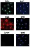
Fig. 1
Fig. 1
Immunostaining demonstrate that our OPC cultures express the expected markers A2B5 (green) and NG2 (red). The lack of GFAP (red) show that these cultures are not contaminated by astrocytes. Nuclei are stained with DAPI (blue).
Fig. 2
Fig. 2
Representative images of OPC cultures show that H2O2 treatment (10 μM for 24 hr) induce cell damage in OPCs. Co-treatment with astrocyte-conditioned media (Astro-CM) blocks the H2O2-induced cell death.
Fig. 3
Fig. 3
(A) A cell survival (WST) assay shows that H2O2 treatment induces OPC death in a concentration-dependent manner. Astrocyte-conditioned media (Astro-CM) are OPC-supportive both under normal and stressed conditions. The assay was conducted 24-hr after H2O2 and/or Astro-CM treatment (see Materials and Methods). ☆P < 0.05 vs Control Media in each H2O2 condition. (B) The OPC survival from 3A were recalculated by normalizing against baseline of Control media or Astro-CM alone without H2O2 injury. These renormalized data suggest that Astro-CM may have both “protective” and “proliferative” effects in OPC cultures. ☆P < 0.05 vs Control Media in each H2O2 condition. (C) Conditioned media from NIH-3T3 cells (mouse fibroblast) do not promote OPC proliferation nor protect OPCs against H2O2-induced stress. NIH-3T3-conditioned media were prepared using the same procedures as Astro-CM.
Fig. 4
Fig. 4
(A) Removing supplements from OPC culture media causes OPC death in a time-dependent manner. Conditioned media from astrocyte (Astro-CM) support the OPC survival under starvation conditions. ☆P < 0.05 vs Control Media in each starvation condition. (B) Astro-CM also show OPC-protective effects against oxygen-glucose deprivation (OGD)-induced stress. ☆P < 0.05.
Fig. 5
Fig. 5
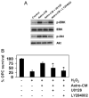
(A) Conditioned media from astrocytes (Astro-CM, 20 min treatment) increase phospho-ERK levels and phospho-Akt levels in recipient OPCs. A specific MEK inhibitor U0126 and a specific PI3K inhibitor LY294002 blocks Astro-CM-induced ERK phosphorylation and Akt phosphorylation, respectively. U0126 (10 μM) and LY294002 (10 μM) were added to OPCs 30 min before Astro-CM treatment. (B) Both U0126 and LY294002 block the ability of Astro-CM to protect OPCs against H2O2 (10 μM)-induced cell damage. ☆P < 0.05 vs H2O2 & Astro-CM.

References

    1. Abbott NJ, Ronnback L, Hansson E. Astrocyte-endothelial interactions at the blood-brain barrier. Nat Rev Neurosci. 2006;7:41–53. - PubMed
    1. Arai K, Lee SR, Lo EH. Essential role for ERK mitogen-activated protein kinase in matrix metalloproteinase-9 regulation in rat cortical astrocytes. Glia. 2003;43:254–264. - PubMed
    1. Arai K, Lo EH. An oligovascular niche: cerebral endothelial cells promote the survival and proliferation of oligodendrocyte precursor cells. J Neurosci. 2009;29:4351–4355. - PMC - PubMed
    1. Butt AM, Ibrahim M, Ruge FM, Berry M. Biochemical subtypes of oligodendrocyte in the anterior medullary velum of the rat as revealed by the monoclonal antibody Rip. Glia. 1995;14:185–197. - PubMed
    1. Canoll PD, Musacchio JM, Hardy R, Reynolds R, Marchionni MA, Salzer JL. GGF/neuregulin is a neuronal signal that promotes the proliferation and survival and inhibits the differentiation of oligoden-drocyte progenitors. Neuron. 1996;17:229–243. - PubMed

Publication types

MeSH terms