. 2010 Mar;88(4):917-26.
doi: 10.1002/jnr.22252.

Fear learning and extinction are linked to neuronal plasticity through Rin1 signaling

Affiliations

Fear learning and extinction are linked to neuronal plasticity through Rin1 signaling

Joanne M Bliss et al. J Neurosci Res. 2010 Mar.

Abstract

The amygdala is known to have a crucial role in both the acquisition and extinction of conditioned fear, but the physiological changes and biochemical mechanisms underlying these forms of learning are only partly understood. The Ras effector Rin1 activates Abl tyrosine kinases and Rab5 GTPases and is highly expressed in mature neurons of the telencephalon including the amygdala, where it inhibits the acquisition of fear memories (Rin1(-/-) mice show enhanced learning of conditioned fear). Here we report that Rin1(-/-) mice exhibit profound deficits in both latent inhibition and fear extinction, suggesting a critical role for Rin1 in gating the acquisition and persistence of cue-dependent fear conditioning. Surprisingly, we also find that depotentiation, a proposed cellular mechanism of extinction, is enhanced at lateral-basolateral (LA-BLA) amygdaloid synapses in Rin1(-/-) mice. Inhibition of a single Rin1 downstream effector pathway, the Abl tyrosine kinases, led to reduced amygdaloid depotentiation, arguing that proper coordination of Abl and Rab5 pathways is critical for Rin1-mediated effects on plasticity. While demonstrating a correlation between amygdala plasticity and fear learning, our findings argue against models proposing a direct causative relationship between amygdala depotentiation and fear extinction. Taken together, the behavior and physiology of Rin1(-/-) mice provide new insights into the regulation of memory acquisition and maintenance. In addition, Rin1(-/-) mice should prove useful as a model for pathologies marked by enhanced fear acquisition and retention, such as posttraumatic stress disorder.


Figures

Fig. 1
Fig. 1
Fear Extinction is reduced in Rin1−/− mice. A. Outline of extinction protocol used. B. WT mice that have undergone extinction freeze less than controls during a tone test (WT ctrl., n = 17 mice; WT extn., n = 17 mice) *p < 0.05. C. Rin1−/− mice show no extinction with equal freezing in control and extinguished mice (Rin1−/− ctrl., n = 17 mice; Rin1−/− extn., n = 17 mice). PRE-CS = baseline freezing before CS presentation; CS = freezing during CS presentation.
Fig. 2
Fig. 2
Rin1−/− mice are deficient in latent inhibition. A. Outline of latent inhibition protocol used. B. WT mice show latent inhibition of fear, with less freezing in preexposed (prx) mice compared to non-preexposed controls (WT non-prx, n = 10 mice; WT prx, n = 10 mice) *p < 0.05. C. Rin1−/− mice display little to no latent inhibition (Rin1−/− non-prx, n = 10 mice; Rin1−/− prx, n = 10 mice). PRE-CS = baseline freezing before CS presentation; CS = freezing during CS presentation; LI = latent inhibition.
Fig. 3
Fig. 3
Rin1−/− mice show normal levels of prepulse inhibition. Levels of startle were measured for pulses (p) of 105 or 120 dB with or without prepulses (pp) of 68, 71, or 77 dB. Prepulse inhibition (PPI) is calculated as [1-(startle to prepulse with pulse/startle to pulse alone)], i.e. a decrease in startle after the prepulse translates to greater PPI. Rin1−/− mice were not significantly altered from WT mice at any of the prepulse or pulse levels. (WT, n = 13 mice; Rin1−/−, n = 18 mice).
Fig. 4
Fig. 4
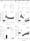
Depotentiation at LA-BLA synapses is enhanced in Rin1−/− mice. A. High-frequency presynaptic fiber stimulation (HFS; 5 trains of 100 Hz stimulation, 1 sec in duration) induced similar levels of LTP in slices from wild type (open symbols, n = 5 slices from 5 mice) and Rin1−/− mice (filled symbols, n = 5 slices from 5 mice). The inset shows superimposed postsynaptic responses recorded during baseline and 60 min post-HFS in slices from wild type (left) and Rin1−/− mice. Calibration bars are 5.0 milliseconds and 0.5 mV. B. Low-frequency stimulation (LFS, 1 Hz for 15 min.) delivered starting 20 min after the last train of HFS induced stronger depotentiation in slices from Rin1−/− mice (filled symbols, n = 7 slices from 4 mice) compared to wild-type mice (open symbols, n = 8 slices from 4 mice). Over the last 10 min of the experiment PSPs in wild type slices were significantly larger that those seen in slices from Rin1−/− mice (p < 0.05). The inset shows postsynaptic responses recorded during baseline (left), 20 min post-HFS (middle), and 45 min post-LFS (right) in slices from wild type (top) and Rin1−/− mice (bottom). Calibration bars are 5.0 milliseconds and 0.5 mV. C. Summary of the effects of HFS and HFS followed by LFS in slices from wild type and Rin1−/− mice. Values correspond to normalized PSP amplitudes recorded 80 min post-HFS in control LTP experiments (open bars) and in experiments where LFS was delivered post-HFS (filled bars) shown in panels A and B. Note that while LFS had no significant effect on LTP in wild type slices it induced significant depotentiation in Rin1−/− slices. D. Fifteen min of 1 Hz stimulation had little lasting effect on LA-BLA synaptic transmission in slices from wild type (open symbols, n = 14 slices from 7 mice) and Rin1−/− mice (filled symbols, n = 20 slices from 7 mice). Although LFS appears to induce larger LTD in slices from Rin1−/− mice, this difference was not statistically significant.
Fig. 5
Fig. 5
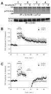
The ABL tyrosine kinase inhibitor imatinib inhibits depotentiation at LA-BLA synaspes. A. Forebrain slices from four mice were pre-incubated with 5 μM imatinib or drug free ACSF for one hr before electrophysiology. Subsequently, slice extracts were analyzed by immunoprecipitation and immunoblot for changes in tyrosine phosphorylation of endogenous Crkl, an Abl substrate. MW markers (kDa) are shown at left. The constant 55 kDa band corresponds to antibody heavy chain. B. HFS induced similar levels of LTP in imatinib-treated (filled symbols, n = 6 slices from 6 mice) and control untreated (open symbols, n = 5 slices from 5 mice) samples. C. LFS delivered 15 min post-HFS induced strong depotentiation in control experiments (open symbols, n = 7 slices from 7 mice) but induced significantly less depotentiation in slices bathed in ACSF containing 5 μM imatinib (filled symbols, n = 4 slices from 4 mice, p < 0.05 compared to untreated controls).
Fig. 6
Fig. 6
LTP, depotentiation and LTD are normal in the hippocampal CA1 region of Rin1−/− mice. A. Two trains of 100 Hz stimulation (HFS) delivered at time = 0 induced similar levels of LTP in slices from wild-type mice (open symbols, n = 8 slices from 7 mice) and Rin1−/− mice (filled symbols, n = 8 slices from 8 mice). The inset shows fEPSPs recorded during baseline and 60 min post-HFS in slices from wild type (left) and Rin1−/− mice (right). Calibration bars are 5.0 milliseconds and 2.0 mV. B. LFS (1 Hz/15min.) delivered 15 min post-HFS induced similar levels of depotentiation in slices from wild-type mice (open symbols, n = 8 slices from 7 mice) and Rin1−/− mutant mice (filled symbols, n = 8 slices from 8 mice). C. The induction of LTD in the hippocampal CA1 region is not enhanced in Rin1−/− mice. Sixty minute post-LFS fEPSPs were depressed to 73 ± 2% of baseline in slices from wild-type mice (open symbols, n = 10 slices from 5 mice) and were depressed to 70 ± 3% of baseline in slices from Rin1−/− mice (filled symbols, n = 8 slices from 4 mice).

References

    1. Anagnostaras SG, Josselyn SA, Frankland PW, Silva AJ. Computer-assisted behavioral assessment of Pavlovian fear conditioning in mice. Learn Mem. 2000;7:58–72. - PMC - PubMed
    1. Barbieri MA, Kong C, Chen PI, Horazdovsky BF, Stahl PD. The SRC homology 2 domain of Rin1 mediates its binding to the epidermal growth factor receptor and regulates receptor endocytosis. J Biol Chem. 2003;278:32027–32036. - PubMed
    1. Bouton ME. Context and behavioral processes in extinction. Learn Mem. 2004;11:485–494. - PubMed
    1. Brown TC, Tran IC, Backos DS, Esteban JA. NMDA receptor-dependent activation of the small GTPase Rab5 drives the removal of synaptic AMPA receptors during hippocampal LTD. Neuron. 2005;45:81–94. - PubMed
    1. Cao X, Tanis KQ, Koleske AJ, Colicelli J. Enhancement of ABL kinase catalytic efficiency by a direct binding regulator is independent of other regulatory mechanisms. J Biol Chem. 2008;283:31401–31407. - PMC - PubMed

Publication types

MeSH terms

Substances