. 2009 Nov 11;131(44):16096-107.
doi: 10.1021/ja902052v.

Structural perturbations induced by the alpha-anomer of the aflatoxin B(1) formamidopyrimidine adduct in duplex and single-strand DNA

Affiliations
Free PMC article

Structural perturbations induced by the alpha-anomer of the aflatoxin B(1) formamidopyrimidine adduct in duplex and single-strand DNA

Kyle L Brown et al. J Am Chem Soc. .
Free PMC article

Abstract

The guanine N7 adduct of aflatoxin B(1) exo-8,9-epoxide hydrolyzes to form the formamidopyrimidine (AFB-FAPY) adduct, which interconverts between alpha and beta anomers. The beta anomer is highly mutagenic in Escherichia coli, producing G --> T transversions; it thermally stabilizes the DNA duplex. The AFB-alpha-FAPY adduct blocks replication; it destabilizes the DNA duplex. Herein, the structure of the AFB-alpha-FAPY adduct has been elucidated in 5'-d(C(1)T(2)A(3)T(4)X(5)A(6)T(7)T(8)C(9)A(10))-3'.5'-d(T(11)G(12)A(13)A(14)T(15)C(16)A(17)T(18)A(19)G(20))-3' (X = AFB-alpha-FAPY) using molecular dynamics calculations restrained by NMR-derived distances and torsion angles. The AFB moiety intercalates on the 5' face of the pyrimidine moiety at the damaged nucleotide between base pairs T(4).A(17) and X(5).C(16), placing the FAPY C5-N(5) bond in the R(a) axial conformation. Large perturbations of the epsilon and zeta backbone torsion angles are observed, and the base stacking register of the duplex is perturbed. The deoxyribose orientation shifts to become parallel to the FAPY base and displaced toward the minor groove. Intrastrand stacking between the AFB moiety and the 5' neighbor thymine remains, but strong interstrand stacking is not observed. A hydrogen bond between the formyl group and the exocyclic amine of the 3'-neighbor adenine stabilizes the E conformation of the formamide moiety. NMR studies reveal a similar 5'-intercalation of the AFB moiety for the AFB-alpha-FAPY adduct in the tetramer 5'-d(C(1)T(2)X(3)A(4))-3', involving the R(a) axial conformation of the FAPY C5-N(5) bond and the E conformation of the formamide moiety. Since in duplex DNA the AFB moiety of the AFB-beta-FAPY adduct also intercalates on the 5' side of the pyrimidine moiety at the damaged nucleotide, we conclude that favorable 5'-stacking leads to the R(a) conformational preference about the C5-N(5) bond; the same conformational preference about this bond is also observed at the nucleoside and base levels. The structural distortions and the less favorable stacking interactions induced by the AFB-alpha-FAPY adduct explain its lower stability as compared to the AFB-beta-FAPY adduct in duplex DNA. In this DNA sequence, hydrogen bonding between the formyl oxygen and the exocyclic amine of the 3'-neighboring adenine stabilizing the E configuration of the formamide moiety is also observed for the AFB-beta-FAPY adduct, and suggests that the identity of the 3'-neighbor nucleotide modulates the stability and biological processing of AFB adducts.


Figures

Scheme 1
Scheme 1. Chemistry of the FAPY Adduct Following Base-Catalyzed Ring-Opening of the AFB-N7-dGuo Cationic Adduct
The adduct interconverts between AFB-α-FAPY and AFB-β-FAPY anomers; the equilibrium is dependent on single strand vs duplex DNA environments.(32) Geometrical isomers of the formamide group and atropisomers about the C5−N5 bond are also observed.(32)
Figure 1
Figure 1
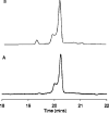
HPLC analyses of the AFB-α-FAPY modified duplex, monitored by UV absorbance at 360 nm. (A) Freshly prepared sample. (B) Three days after preparing the sample. The oligodeoxynucleotide containing the AFB-α-FAPY adduct eluted at 20.1 min. The oligodeoxynucleotide containing the AFB-β-FAPY adduct eluted at 19.5 min.
Figure 2
Figure 2
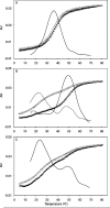
Thermal denaturation studies monitored by UV absorbance at 260 nm showing heating (▲) and cooling (Δ) transitions. (A) The unmodified duplex 5′-d(CTATGATTCA)-3′·5′-d(TGAATCATAG)-3′. (B) The duplex containing the AFB-β-FAPY adduct. (C) The duplex containing the AFB-α-FAPY adduct. The solid lines are first derivative curves.
Figure 3
Figure 3
Analysis of NOE intensities for the deoxyribose protons. (A) 5′-d(CTATXATTCA)-3′·5′-d(TGAATCATAG)-3′. (B) 5′-d(CTXA)-3′, X = AFB-α-FAPY.
Figure 4
Figure 4
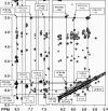
A tile plot of an 800 MHz NOESY spectrum obtained at 250 ms mixing time, showing the assignments of AFB-α-FAPY protons and NOEs to neighboring nucleotide protons. The black lines indicate the assignmnents of the AFB-α-FAPY protons and the gray lines indicate NOEs between the AFB moiety and DNA.
Figure 5
Figure 5
Chemical shift perturbation of AFB-α-FAPY modified duplex relative to the unmodified oligodeoxynucleotide. (A) The aromatic H6/H8 (black bars) and pyrimidine H5/CH3 (white bars) resonances of the modified strand. (B) The aromatic H6/H8 (black bars) and pyrimidine H5/CH3 (white bars) resonances of the complementary strand. (C) The deoxyribose resonances H1′ (black bars), H2′ (white bars), H2′′ (dark gray bars), and H3′ (light gray bars) of the modified strand. (D) The deoxyribose resonances H1′ (black bars), H2′ (white bars), H2′′ (dark gray bars), and H3′ (light gray bars) of the complementary strand. (E) The exchangeable N1H/N3H (black bars) resonances. (F) Comparison of deoxyribose resonances H1′ (black bars), H2′ (white bars), H2′′ (dark gray bars), and H3′ (light gray bars) for corresponding nucleotides of the duplex and single strand tetramer.
Figure 6
Figure 6
Stereoview of nine superimposed structures of the AFB-α-FAPY modified duplex extracted from isothermal rMD calculations in explicit solvent (PDB ID: 2KH3). The AFB moiety and the formyl oxygen are depicted in red. The R1x residual of 8.7 × 10⁻² for the ensemble indicates agreement with NOESY data (Table 3).
Figure 7
Figure 7
(A) Refined structure of the AFB-β-FAPY adduct in duplex DNA (PDB ID: 1HM1).(38) (B) Refined structure of the AFB-α-FAPY adduct in duplex DNA (PDB ID: 2KH3). (C) Molecular model of the AFB-α-FAPY adduct the single stranded tetramer (PDB ID: 2KH4).
Figure 8
Figure 8
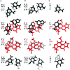
Stacking interactions of AFB-α-FAPY adduct in single-strand (molecular modeling) and duplex DNA (refined structure) compared with the stacking interactions of the AFB-β-FAPY adduct (refined structure) in duplex DNA. (A) The AFB-β-FAPY adduct in duplex DNA (PDB ID: 1HM1).(38) (B) The AFB-α-FAPY adduct in duplex (PDB ID: 2KH3). (C) The AFB-α-FAPY adduct as modeled in the tetramer 5′-d(CTXA)-3′ (PDB ID: 2KH4).
Figure 9
Figure 9
Overlay of AFB-α-FAPY and AFB-β-FAPY adducts, showing differences in phosphodiester backbone geometry. AFB-β-FAPY is depicted with light gray carbon atoms and green hydrogen atoms. AFB-α-FAPY is depicted with dark gray carbon atoms and white hydrogen atoms. In both structures, oxygen atoms are red and phosphorus atoms are orange.

Similar articles

Cited by

References

    1. Busby W. F. Jr.; Wogan G. N. In. Chemical Carcinogens; 2nd ed.; Searle C. E., Ed.; American Chemical Society: Washington, D.C., 1984; pp 945−1136.
    1. Smela M. E.; Currier S. S.; Bailey E. A.; Essigmann J. M. Carcinogenesis 2001, 22, 535–545. - PubMed
    1. Bennett J. W.; Klich M. Clin. Microbiol. Rev. 2003, 16, 497–516. - PMC - PubMed
    1. McCann J.; Spingarn N. E.; Koburi J.; Ames B. N. Proc. Natl. Acad. Sci. U.S.A. 1975, 72, 979–983. - PMC - PubMed
    1. Foster P. L.; Eisenstadt E.; Miller J. H. Proc. Natl. Acad. Sci. U.S.A. 1983, 80, 2695–2698. - PMC - PubMed

Publication types