

Vitreous metastases of primary cutaneous B-cell lymphoma
- PMID: 19831568
- PMCID: PMC2773692
- DOI: 10.3109/09273940903144705
Vitreous metastases of primary cutaneous B-cell lymphoma
Abstract
Purpose: To describe two cases of vitreous metastases of primary cutaneous B-cell lymphoma (PCBCL).
Methods: Observational case series.
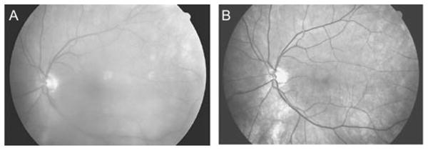
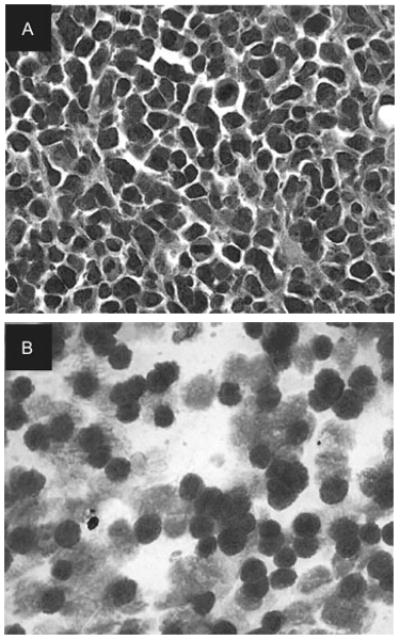
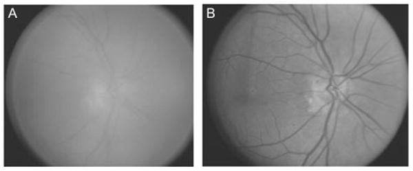
Results: A 73-year-old man and an 81-year-old woman, both with a history of PCBCL, diffuse large cell type, presented with decreased visual acuity due to vitritis. Both patients underwent vitreous biopsy that demonstrated B-cell lymphoma, large cell type, and confirmed metastases of cutaneous B-cell lymphoma to the vitreous.
Conclusion: PCBCL, diffuse large cell type, is a rare form of non-Hodgkin lymphoma that can metastasize to the vitreous without visible chorioretinal involvement.
Conflict of interest statement
Figures
References
-
- Groves FD, Linet MS, Travis LB, Devesa SS. Cancer surveillance series: non-Hodgkin's lymphoma incidence by histologic subtype in the United States from 1978 through 1995. J Natl Cancer Inst. 2000;92(15):1240–1251. - PubMed
-
- Willemze R, Jaffe ES, Burg G, et al. WHO-EORTC classification for cutaneous lymphomas. Blood. 2005;105(10):3768–3785. - PubMed
-
- Grange F, Beylot-Barry M, Courville P, et al. Primary cutaneous diffuse large B-cell lymphoma, leg type: clinicopathologic features and prognostic analysis in 60 cases. Arch Dermatol. 2007;143(9):1144–1150. - PubMed
-
- Chan CC, Gonzales JA. Primary Intraocular Lymphoma Singapore. London, Hackensack NJ: World Scientific Publishing Co.; 2007.
Publication types
MeSH terms
Grants and funding
LinkOut - more resources
Full Text Sources
Medical