. 2009 Oct 16;16(1):94.
doi: 10.1186/1423-0127-16-94.

Antinociceptive actions of honokiol and magnolol on glutamatergic and inflammatory pain

Affiliations

Antinociceptive actions of honokiol and magnolol on glutamatergic and inflammatory pain

Yi-Ruu Lin et al. J Biomed Sci. .

Abstract

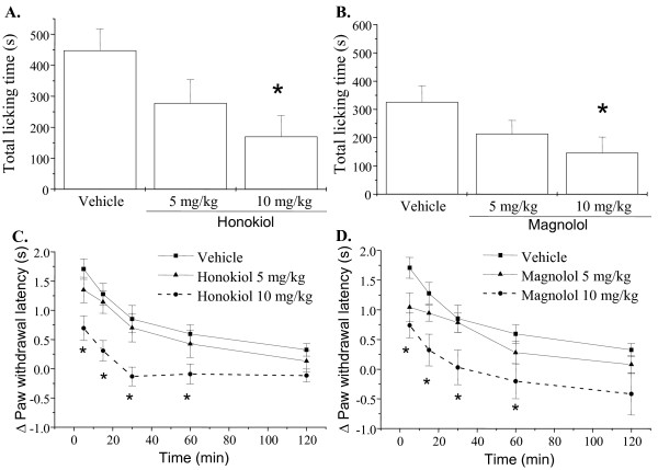
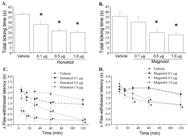
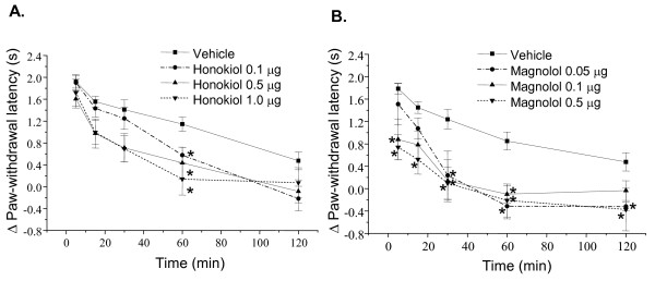
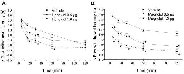
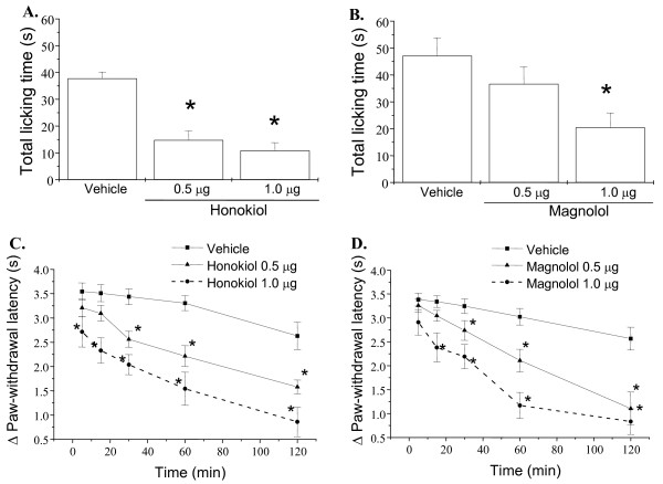
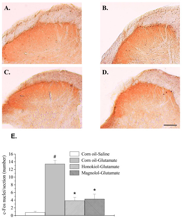
The antinociceptive effects of honokiol and magnolol, two major bioactive constituents of the bark of Magnolia officinalis, were investigated on animal paw licking responses and thermal hyperalgesia induced by glutamate receptor agonists including glutamate, N-methyl-D-aspartate (NMDA), and metabotropic glutamate 5 receptor (mGluR5) activator (RS)-2-chloro-5-hydroxyphenylglycine (CHPG), as well as inflammatory mediators such as substance P and prostaglandin E2 (PGE2) in mice. The actions of honokiol and magnolol on glutamate-induced c-Fos expression in the spinal cord dorsal horn were also examined. Our data showed that honokiol and magnolol blocked glutamate-, substance P- and PGE2-induced inflammatory pain with similar potency and efficacy. Consistently, honokiol and magnolol significantly decreased glutamate-induced c-Fos protein expression in superficial (I-II) laminae of the L4-L5 lumbar dorsal horn. However, honokiol was more selective than magnolol for inhibition of NMDA-induced licking behavioral and thermal hyperalgesia. In contrast, magnolol was more potent to block CHPG-mediated thermal hyperalgesia. These results demonstrate that honokiol and magnolol effectively decreased the inflammatory pain. Furthermore, their different potency on inhibition of nociception provoked by NMDA receptor and mGluR5 activation should be considered.


Figures

Figure 1
Figure 1
Effects of honokiol and magnolol on glutamate-induced nociceptive responses and thermal hyperalgesia in mice. Honokiol (5 and 10 mg/kg), magnolol (5 and 10 mg/kg) and vehicle (corn oil) were intraperitoneally administered 20 min prior to glutamate (300 μg) injection into hindpaw, and the total time spent licking the hindpaw was measured for 30 min after intraplantar injection of glutamate (A, B). The paw withdrawal latency in hot water at 47°C was measured during 5-120 min after intraplantar injection of glutamate (C, D). Each point represents the mean ± S.E.M. (n = 6-8). * indicates significantly different from vehicle group, *p < 0.05.
Figure 2
Figure 2
Effects of honokiol and magnolol on NMDA-induced nociceptive responses and thermal hyperalgesia in mice. Honokiol (0.1-1.0 μg/paw), magnolol (0.1-1.0 μg/paw) and vehicle were intraplantarly administered 20 min prior to NMDA (150 μg) injection into hindpaw, and the total time spent licking the hindpaw was measured for 5 min after intraplantar injection of NMDA (A, B). The paw withdrawal latency in hot water at 47°C was measured during 5-120 min after intraplantar injection of NMDA (C, D). Each point represents the mean ± S.E.M. (n = 8-16). * indicates significantly different from vehicle group, *p < 0.05.
Figure 3
Figure 3
Effects of honokiol and magnolol on CHPG-induced thermal hyperalgesia in mice. Honokiol (0.1-1.0 μg/paw), magnolol (0.05-0.5 μg/paw) and vehicle were administered 20 min prior to CHPG (1 μg) injection into hindpaw. The paw withdrawal latency in the 47°C water was measured during 5-120 min after intraplantar injection of CHPG. Each point represents the mean ± S.E.M. (n = 7-19). * indicates significantly different from vehicle group, *p < 0.05.
Figure 4
Figure 4
Effects of honokiol and magnolol on substance P-induced thermal hyperalgesia in mice. Honokiol (0.5 or 1.0 μg/paw), magnolol (0.5 or 1.0 μg/paw) and vehicle were administered 20 min prior to substance-P (0.5 μg) injection into hindpaw. The paw withdrawal latency in the 47°C water was measured during 5-120 min after intraplantar injection of substance P. Each point represents the mean ± S.E.M. (n = 7-9). * indicates significantly different from vehicle group, *p < 0.05.
Figure 5
Figure 5
Effects of honokiol and magnolol on PGE2-induced nociceptive responses and thermal hyperalgesia in mice. Honokiol (0.5 or 1.0 μg/paw), magnolol (0.5 or 1.0 μg/paw) and vehicle were administered 20 min prior to PGE2 (0.5 μg) injection into hindpaw, and the total time spent licking the hindpaw was measured for 5 min after intraplantar injection of PGE2 (A, B). The paw withdrawal latency in the 47°C water was measured during 5-120 min after intraplantar injection of PGE2 (C, D). Each point represents the mean ± S.E.M. (n = 7-8). * indicates significantly different from vehicle group, *p < 0.05.
Figure 6
Figure 6
Effects of honokiol and magnolol on glutamate-induced c-Fos protein expression in the spinal dorsal horn of the side ipsilateral to the injection. Photomicrographs illustrating the four experimental groups are represented: corn oil i.p.+saline i.pl. (A), corn oil i.p.+glutamate i.pl. (B), honokiol (10 mg/kg) i.p.+glutamate i.pl. (C), and magnolol (10 mg/kg) i.p.+glutamate i.pl. (D). Summary data of glutamate-induced c-Fos protein expression after honokiol and magnolol treatment in superficial (I-II) spinal cord (E). Values are means ± S.E.M. (n = 4). # indicates significantly different from corn oil-saline group, #P < 0.05; * indicates significantly different from corn oil-glutamate group, *P < 0.05. Statistical analysis was determined by one-way ANOVA followed by a Student-Newman-Keuls post-hoc test. Scale bar = 100 μm.

References

    1. Akerud P, Canals JM, Snyder EY, Arenas E. Neuroprotection through delivery of glial cell line-derived neurotrophic factor by neural stem cells in a mouse model of Parkinson's disease. J Neurosci. 2001;21:8108–8118. - PMC - PubMed
    1. Otsuka M, Konishi S. Release of substance P-like immunoreactivity from isolated spinal cord of newborn rat. Nature. 1976;264:83–84. doi: 10.1038/264083a0. - DOI - PubMed
    1. McCarson KE, Goldstein BD. Time course of the alteration in dorsal horn substance P levels following formalin: blockade by naloxone. Pain. 1990;41:95–100. doi: 10.1016/0304-3959(90)91113-W. - DOI - PubMed
    1. Broman J, Adahl F. Evidence for vesicular storage of glutamate in primary afferent terminals. Neuroreport. 1994;5:1801–1804. doi: 10.1097/00001756-199409080-00029. - DOI - PubMed
    1. Sluka KA, Willis WD. Increased spinal release of excitatory amino acids following intradermal injection of capsaicin is reduced by a protein kinase G inhibitor. Brain Res. 1998;798:281–286. doi: 10.1016/S0006-8993(98)00428-4. - DOI - PubMed

Publication types

MeSH terms