. 2009 Oct 16;31(4):565-75.
doi: 10.1016/j.immuni.2009.07.010.

Marked induction of the helix-loop-helix protein Id3 promotes the gammadelta T cell fate and renders their functional maturation Notch independent

Affiliations

Marked induction of the helix-loop-helix protein Id3 promotes the gammadelta T cell fate and renders their functional maturation Notch independent

Jens Peter Holst Lauritsen et al. Immunity. .

Abstract

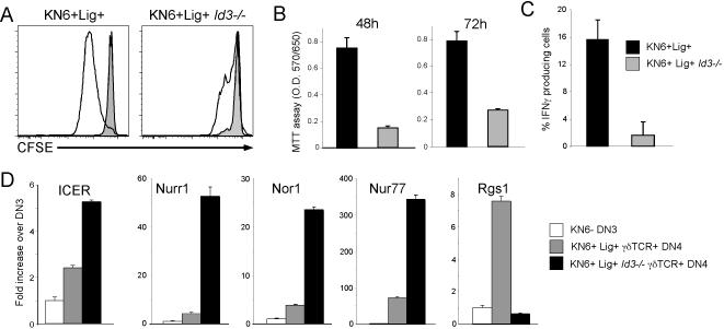
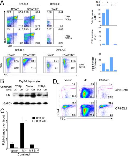
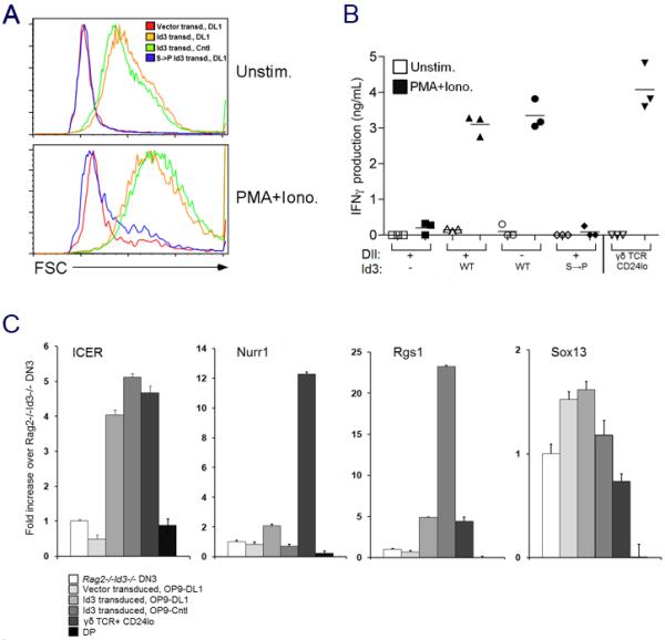
alphabeta and gammadelta T cells arise from a common thymocyte progenitor during development in the thymus. Emerging evidence suggests that the pre-T cell receptor (pre-TCR) and gammadelta T cell receptor (gammadeltaTCR) play instructional roles in specifying the alphabeta and gammadelta T-lineage fates, respectively. Nevertheless, the signaling pathways differentially engaged to specify fate and promote the development of these lineages remain poorly understood. Here, we show that differential activation of the extracellular signal-related kinase (ERK)-early growth response gene (Egr)-inhibitor of DNA binding 3 (Id3) pathway plays a defining role in this process. In particular, Id3 expression served to regulate adoption of the gammadelta fate. Moreover, Id3 was both necessary and sufficient to enable gammadelta-lineage cells to differentiate independently of Notch signaling and become competent IFNgamma-producing effectors. Taken together, these findings identify Id3 as a central player that controls both adoption of the gammadelta fate and its maturation in the thymus.


Figures

Figure 1
Figure 1. Id3 is required for Egr1-mediated promotion of γδ development
(A) Egr1, Egr3, and Id3 mRNA are more highly expressed in developing γδ lineage cells. Egr and Id mRNA levels were quantified in triplicate in the indicated cell populations by Real time PCR, standardized using β-actin, and normalized to levels in DN3. Results ± standard deviation are depicted graphically. (B-C) Induction of γδ development by enforced expression of Egr1 is dependent on Id3. E13.5 Id3+/- and Id3-/- fetal thymocytes were transduced with empty vector (LZRS) or Egr1 (LZRS-Egr1), seeded into thymic lobes and placed in fetal thymic organ culture for 2 days. Developmental progression was assessed by flow cytometry on electronically gated transduced (GFP+) cells. The absolute number of γδ lineage cells (γδTCR+ or γδTCR+CD24lo) and αβ lineage DP thymocytes per lobe is depicted graphically in (C). Results are representative of 3 experiments performed.
Figure 2
Figure 2. Id3-deficiency disrupts the development of γδ T cells
(A-B) Id3-deficiency results in expansion of the Vγ1.1+ subset. Id3-/- mice were backcrossed to the C57BL/6 background for 3 generations before tissues from mice of the indicated genotypes were analyzed by flow cytometry. Results are depicted as histograms and summarized graphically on the right. (C-D) Id3-deficiency rescues deletion of autoreactive γδ precursors. Thymocyte preparations were produced from Rag2+/- KN6 Tg mice expressing positively selecting ligand (T-22d) or higher affinity negatively selecting ligand (T-10/22d/b) and from KN6 Tg mice expressing high affinity ligand but lacking Id3. Preparations were analyzed by flow cytometry and gated DN cells were displayed as histograms as above. Differences in absolute number of the indicated subsets that are marked by asterisks are statistically significant (p<0.05). Results are representative of 3 experiments performed with a minimum of 2 mice per genotype in each experiment.
Figure 3
Figure 3. Egr1 and Id3 can modulate lineage choice without affecting the TCR repertoire
(A) Schematic representation of lineage assignment in the KN6 γδTCR Tg model. (B) Enforced expression of Egr1 diverts KN6 Tg thymocytes to the γδ fate. Thymocytes from mice of the indicated genotypes were analyzed by flow cytometry: 1) Lig-, KN6 Tg Rag2-/-β2M-/-; 2) Lig-Egr1Tg, KN6 Tg Rag2-/-β2M-/- Egr1Tg. Results are depicted as histograms and summarized graphically on the right with each symbol corresponding to an individual mouse. (C) The strong TCR signals that promote adoption of the γδ fate require Id3. Mice of the indicated genotypes were analyzed by flow cytometry: 1) Lig+, KN6 Tg Rag2-/-; 2) Lig+Id3-/-, KN6 Tg Rag2-/-Id3-/-. (B-C) Differences between the absolute number of DP and CD24lo DN thymocytes between these mice are statistically significant (asterisk indicates p<0.05).
Figure 4
Figure 4. Id3 promotes the γδ fate and opposes the αβ fate and these effects are associated with alternations in growth and survival
(A-B) Id3 promotes survival of mature CD24lo γδ lineage cells and impairs growth and survival of CD8ISP and αβ lineage DP. Apoptosis and proliferation of the indicated populations were evaluated flow cytometrically by Annexin staining and BrdU incorporation, respectively. Each symbol represents an individual experiment involving at least 3 animals, with a line connecting the animals with a different genotype contained within a single experiment (upper panels). The difference in % apoptotic cells between Lig+ and Lig+Id3-/- populations was significant (p<0.05) for the following populations: 1) γδTCR+CD24lo; 2) ISP; and 3) DP. The mean percent BrdU incorporation ± standard deviation is presented graphically (bottom panels) with a minimum of 10 mice per condition. *, p<0.05; **, p<0.01. (C) Effects of Id3 on growth and survival are associated with differential regulation of Bcl2 family members. Expression of the indicated genes was measured by real time PCR on thymocyte subsets purified by flow cytometry. Expression levels were standardized using β-actin, and normalized to levels in Rag2-/- DN3 thymocytes. Data are representative of two different experiments.
Figure 5
Figure 5. Induction of Id3 by strong signals during γδ development is required for functional competence
(A-C) Id3-deficiency impairs TCR-dependent proliferation and production of IFNγ. DN thymocytes from KN6+Lig+ and KN6+Lig+Id3-/- mice were labeled with CFSE, stimulated for 48h on plate-bound anti-CD3 Ab (10μg/ml), and analyzed by FACS (A). DN thymocytes from the same mice as in (A) were stimulated as above and cellular expansion was evaluated on triplicate wells by MTT assay. (B). DN thymocytes were stimulated with plate-bound anti-CD3 Ab for 24 hours, following which the proportion of IFNγ producing cells was determined by intracellular staining and flow cytometry. (C) Mean ± standard deviation is presented graphically. (D) Expression of the γδ biased gene profile and functional competence can be separated. Expression of the indicated genes was measured on flow cytometrically isolated cell populations by real time PCR as in Figure 4. Mean ± standard deviation is presented graphically.
Figure 6
Figure 6. Id3 induction by strong signals is necessary and sufficient to confer Notch-independence on γδ lineage cells
(A) Notch independence of γδ lineage cells is impaired by Id3-deficiency. Rag2-/- or Rag2-/-Id3-/- DN3 cells were transduced as indicated, cultured on OP9-DL1 or OP9-Cntl cells for 5 d, and the expression of CD4 and CD8 was analyzed by flow cytometry. Bar graphs on the right show the fold increases (relative to control transduced cultures) in absolute numbers of DP or γδ+ CD24lo DN cells from the indicated transductions, culture conditions and genotype. (B) Notch signals reduce E protein expression. Equal quantities of protein from detergent extracts of cells cultured as in (A) were immunoblotted with the indicated Ab. (C-D) Ectopic expression of Id3 is sufficient to confer Notch independence on TCR- thymocytes. Rag2-/-Id3-/- DN3 cells were transduced as indicated, cultured on OP9-DL1 or OP9-Cntl cells for 6 d, with the fold increases in cellularity over input shown in (C), and the expression of CD24 and forward-light scatter (FSC) analysis by flow cytometry are shown in (D). These data are representative of at least 3-independent experiments.
Figure 7
Figure 7. Ectopic expression of Id3 is able to confer functional competence on TCR-thymocytes
(A) Id3 transduced cells increase in size upon PMA + ionomycin stimulation. (B) Id3 transduction confers competence to produce IFNγ on Rag2/Id3-deficient DN3 cells. (A-B) Sorted CD45+ GFP+ cells from transduced DN3 cells (as indicated) were stimulated for 36 h with PMA/Ionomycin (PMA/Iono) or placed in culture without stimulation (Unstim) and (A) analyzed by flow cytometry to determine cell size by forward-light scatter (FSC). (B) Levels of IFNγ in culture supernatants were quantified by an antibody-capture ELISA. (C) Id3 transduction confers expression of some γδ-biased genes. RNA transcript levels of Rgs1, ICER, Nurr1, and Sox13 were measured by real time PCR as in Fig. 5 from sorted CD45+ GFP+ cells from transduced DN3 cells cultured on OP9-Cntl or OP9-DL1 cells as indicated.

References

    1. Adams EJ, Strop P, Shin S, Chien YH, Garcia KC. An autonomous CDR3delta is sufficient for recognition of the nonclassical MHC class I molecules T10 and T22 by gammadelta T cells. Nature Immunology. 2008;9:777–784. - PMC - PubMed
    1. Agata Y, Tamaki N, Sakamoto S, Ikawa T, Masuda K, Kawamoto H, Murre C. Regulation of T cell receptor beta gene rearrangements and allelic exclusion by the helix-loop-helix protein, E47. Immunity. 2007;27:871–884. - PubMed
    1. Bain G, Romanow WJ, Albers K, Havran WL, Murre C. Positive and negative regulation of V(D)J recombination by the E2A proteins. Journal of Experimental Medicine. 1999;189:289–300. - PMC - PubMed
    1. Barndt RJ, Dai M, Zhuang Y. Functions of E2A-HEB heterodimers in T-cell development revealed by a dominant negative mutation of HEB. Mol Cell Biol. 2000;20:6677–6685. - PMC - PubMed
    1. Carleton M, Haks MC, Smeele SA, Jones A, Belkowski SM, Berger MA, Linsley P, Kruisbeek AM, Wiest DL. Early growth response transcription factors are required for development of CD4(-)CD8(-) thymocytes to the CD4(+)CD8(+) stage. Journal of Immunology. 2002;168:1649–1658. - PubMed

Publication types

MeSH terms

Substances