

Noninvasive quantification and optimization of acute cell retention by in vivo positron emission tomography after intramyocardial cardiac-derived stem cell delivery
- PMID: 19833262
- PMCID: PMC2803039
- DOI: 10.1016/j.jacc.2009.04.097
Noninvasive quantification and optimization of acute cell retention by in vivo positron emission tomography after intramyocardial cardiac-derived stem cell delivery
Abstract
Objectives: The aim of this study was to quantify acute myocardial retention of cardiac-derived stem cells (CDCs) and evaluate different delivery methods with positron emission tomography (PET).
Background: Success of stem cell transplantation for cardiac regeneration is partially limited by low retention/engraftment of the delivered cells. A clinically applicable method for accurate quantification of cell retention would enable optimization of cell delivery.
Methods: The CDCs were derived from syngeneic, male Wistar Kyoto (WK) rats labeled with [(18)F]-fluoro-deoxy-glucose ((18)FDG) and injected intramyocardially into the ischemic region of female WK rats after permanent left coronary artery ligation. The effects of fibrin glue (FG), bradycardia (adenosine), and cardiac arrest were examined. Imaging with (18)FDG PET was performed for quantification of cell retention. Quantitative polymerase chain reaction (PCR) for the male-specific SRY gene was performed to validate the PET results.
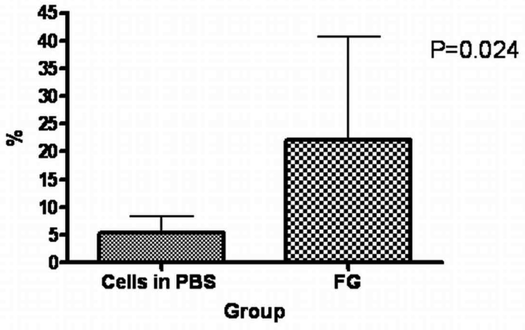
Results: Myocardial retention of cells suspended in phosphate-buffered saline 1 h after delivery was 17.6 +/- 11.5% by PCR and 17.8 +/- 7.3% by PET. When CDCs were injected immediately after induction of cardiac arrest, retention was increased to 75.6 +/- 18.6%. Adenosine slowed the ventricular rate and doubled CDC retention (35.4 +/- 5.3%). A similar increase in CDC retention was observed after epicardial application of FG at the injection site (37.5 +/- 8.2%). The PCR revealed a significant increase in 3-week cell engraftment in the FG animals (22.1 +/- 18.6% and 5.3 +/- 3.1%, for FG and phosphate-buffered saline, respectively).
Conclusions: In vivo PET permits accurate measurement of CDC retention early after intramyocardial delivery. Sealing injection sites with FG or lowering ventricular rate by adenosine might be clinically translatable methods for improving stem cell engraftment in a beating heart.
Conflict of interest statement
No conflicts of interest to disclose for other authors.
Figures


Comment in
-
Tracking cell fate with noninvasive imaging.J Am Coll Cardiol. 2009 Oct 20;54(17):1627-8. doi: 10.1016/j.jacc.2009.05.067. J Am Coll Cardiol. 2009. PMID: 19833263 Free PMC article. No abstract available.
References
-
- Wollert KC, Drexler H. Clinical applications of stem cells for the heart. Circ Res. 2005;96:151–163. - PubMed
-
- Murry CE, Reinecke H, Pabon LM. Regeneration gaps: observations on stem cells and cardiac repair. J Am Coll Cardiol. 2006;47:1777–1785. - PubMed
-
- Segers VF, Lee RT. Stem-cell therapy for cardiac disease. Nature. 2008;451:937–942. - PubMed
-
- Fukushima S, Varela-Carver A, Coppen SR, et al. Direct intramyocardial but not intracoronary injection of bone marrow cells induces ventricular arrhythmias in a rat chronic ischemic heart failure model. Circulation. 2007;115:2254–2261. - PubMed
Publication types
MeSH terms
Grants and funding
LinkOut - more resources
Full Text Sources
Other Literature Sources
