

18F-4V for PET-CT imaging of VCAM-1 expression in atherosclerosis
- PMID: 19833312
- PMCID: PMC2773129
- DOI: 10.1016/j.jcmg.2009.04.016
18F-4V for PET-CT imaging of VCAM-1 expression in atherosclerosis
Abstract
Objectives: The aim of this study was to iteratively develop and validate an (18)F-labeled small vascular cell adhesion molecule (VCAM)-1 affinity ligand and demonstrate the feasibility of imaging VCAM-1 expression by positron emission tomography-computed tomography (PET-CT) in murine atherosclerotic arteries.
Background: Hybrid PET-CT imaging allows simultaneous assessment of atherosclerotic lesion morphology (CT) and may facilitate early risk assessment in individual patients. The early induction, confinement of expression to atherosclerotic lesions, and accessible position in proximity to the blood pool render the adhesion molecule VCAM-1 an attractive imaging biomarker for inflamed atheroma prone to complication.
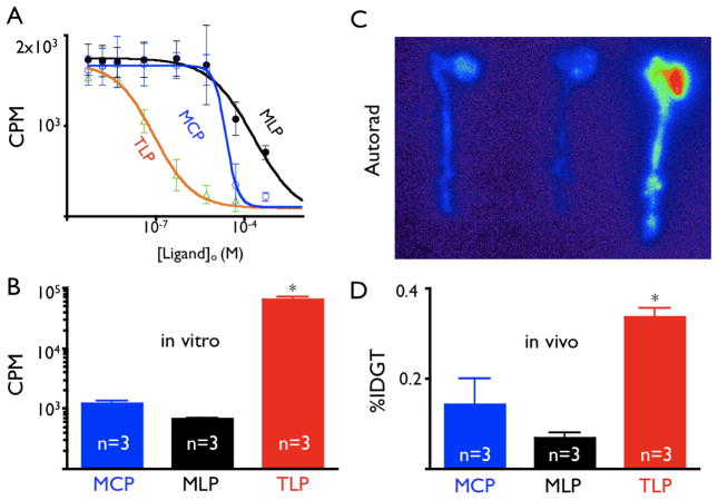
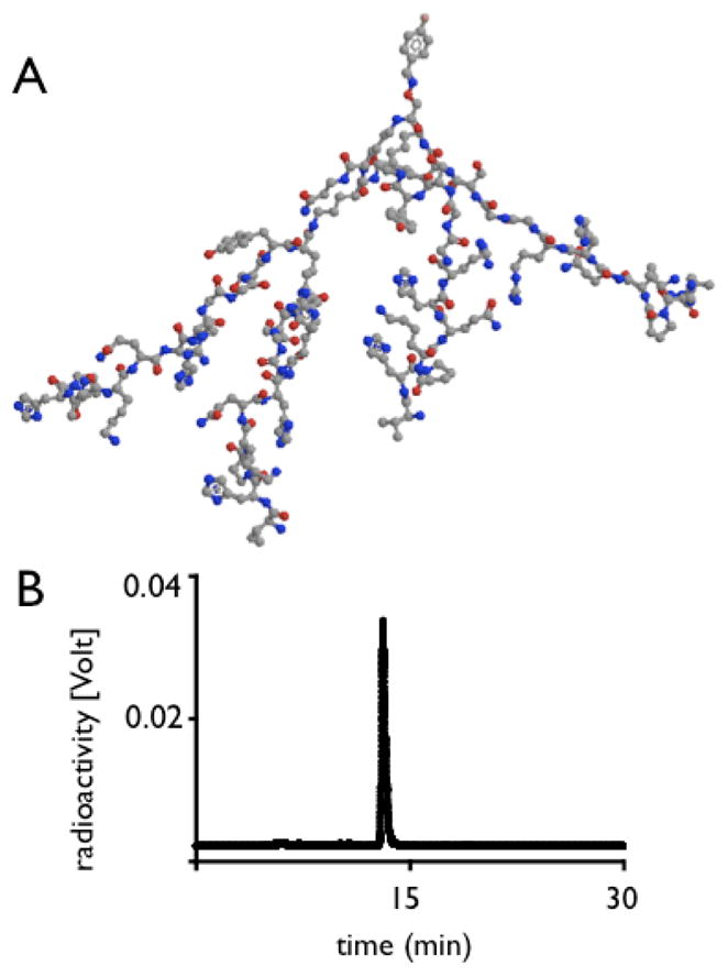
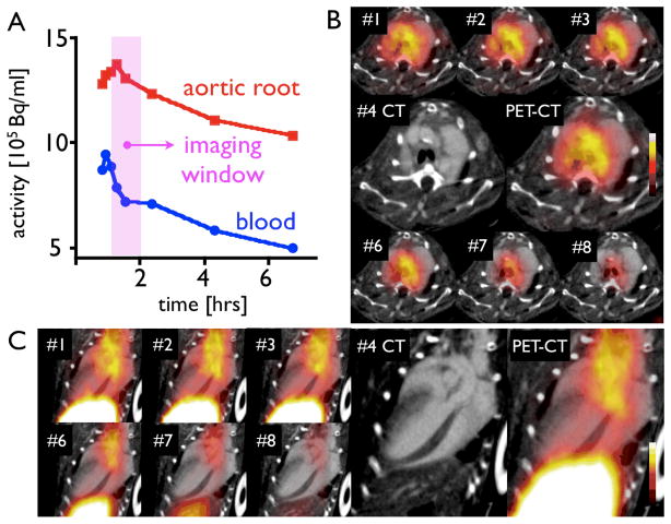
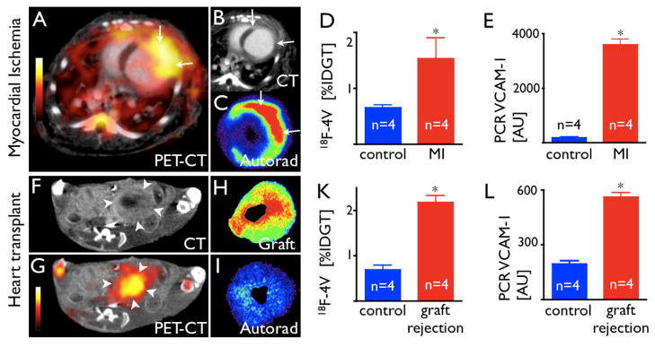
Methods: A cyclic, a linear, and an oligomer affinity peptide, internalized into endothelial cells by VCAM-1-mediated binding, were initially derivatized with DOTA to determine their binding profiles and pharmacokinetics. The lead compound was then (18)F-labeled and tested in atherosclerotic apoE(-/-) mice receiving a high-cholesterol diet as well as wild type murine models of myocardial infarction and heart transplant rejection.
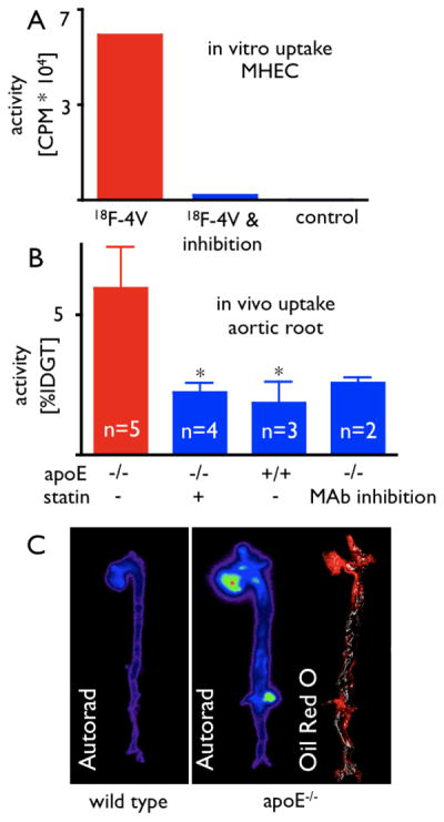
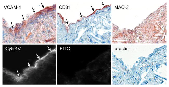
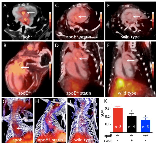
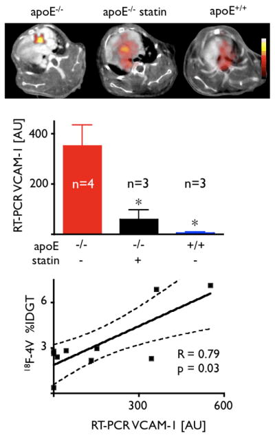
Results: The tetrameric peptide had the highest affinity and specificity for VCAM-1 (97% inhibition with soluble VCAM-1 in vitro). In vivo PET-CT imaging using (18)F-4V showed 0.31 +/- 0.02 SUV in murine atheroma (ex vivo %IDGT 5.9 +/- 1.5). (18)F-4V uptake colocalized with atherosclerotic plaques on Oil Red O staining and correlated to mRNA levels of VCAM-1 measured by quantitative reverse transcription polymerase chain reaction (R = 0.79, p = 0.03). Atherosclerotic mice receiving an atorvastatin-enriched diet had significantly lower lesional uptake (p < 0.05). Furthermore, (18)F-4V imaging in myocardial ischemia after coronary ligation and in transplanted cardiac allografts undergoing rejection showed high in vivo PET signal in inflamed myocardium and good correlation with ex vivo measurement of VCAM-1 mRNA by quantitative polymerase chain reaction.
Conclusions: (18)F-4V allows noninvasive PET-CT imaging of VCAM-1 in inflammatory atherosclerosis, has the dynamic range to quantify treatment effects, and correlates with inflammatory gene expression.
Conflict of interest statement
Conflict of Interest Disclosures: None.
Figures
Comment in
-
Imaging inflammation in atherosclerosis another step forward.JACC Cardiovasc Imaging. 2009 Oct;2(10):1223-5. doi: 10.1016/j.jcmg.2009.06.010. JACC Cardiovasc Imaging. 2009. PMID: 19833313 No abstract available.
Comment on
-
Putting the face to a name: concurrent assessment of vascular morphology and biology.JACC Cardiovasc Imaging. 2009 Oct;2(10):1243-4. doi: 10.1016/j.jcmg.2009.08.005. JACC Cardiovasc Imaging. 2009. PMID: 19833316 No abstract available.
Similar articles
-
Targeting of vascular cell adhesion molecule-1 by 18F-labelled nanobodies for PET/CT imaging of inflamed atherosclerotic plaques.Eur Heart J Cardiovasc Imaging. 2016 Sep;17(9):1001-8. doi: 10.1093/ehjci/jev346. Epub 2016 Jan 22. Eur Heart J Cardiovasc Imaging. 2016. PMID: 26800768
-
Effect of atorvastatin on the expression of gamma-glutamyl transferase in aortic atherosclerotic plaques of apolipoprotein E-knockout mice.BMC Cardiovasc Disord. 2014 Oct 18;14:145. doi: 10.1186/1471-2261-14-145. BMC Cardiovasc Disord. 2014. PMID: 25326709 Free PMC article.
-
Imaging inflammation in atherosclerosis another step forward.JACC Cardiovasc Imaging. 2009 Oct;2(10):1223-5. doi: 10.1016/j.jcmg.2009.06.010. JACC Cardiovasc Imaging. 2009. PMID: 19833313 No abstract available.
-
Variability and uncertainty of 18F-FDG PET imaging protocols for assessing inflammation in atherosclerosis: suggestions for improvement.J Nucl Med. 2015 Apr;56(4):552-9. doi: 10.2967/jnumed.114.142596. Epub 2015 Feb 26. J Nucl Med. 2015. PMID: 25722452 Review.
-
Novel non-invasive approach for visualizing inflamed atherosclerotic plaques using fluorodeoxyglucose-positron emission tomography.Geriatr Gerontol Int. 2010 Jan;10(1):1-8. doi: 10.1111/j.1447-0594.2009.00564.x. Geriatr Gerontol Int. 2010. PMID: 20102376 Review.
Cited by
-
Magnetic resonance coronary angiography: where are we today?Curr Cardiol Rep. 2013 Feb;15(2):328. doi: 10.1007/s11886-012-0328-0. Curr Cardiol Rep. 2013. PMID: 23307168 Free PMC article. Review.
-
Nuclear imaging of inflammation: homing-associated molecules as targets.EJNMMI Res. 2013 Jan 3;3(1):1. doi: 10.1186/2191-219X-3-1. EJNMMI Res. 2013. PMID: 23281702 Free PMC article.
-
Site-Specific Glycation and Chemo-enzymatic Antibody Sortagging for the Retargeting of rAAV6 to Inflamed Endothelium.Mol Ther Methods Clin Dev. 2019 Jul 23;14:261-269. doi: 10.1016/j.omtm.2019.07.003. eCollection 2019 Sep 13. Mol Ther Methods Clin Dev. 2019. PMID: 31453264 Free PMC article.
-
The Role of Molecular Imaging as a Marker of Remyelination and Repair in Multiple Sclerosis.Int J Mol Sci. 2021 Dec 31;23(1):474. doi: 10.3390/ijms23010474. Int J Mol Sci. 2021. PMID: 35008899 Free PMC article. Review.
-
Positron emission tomography imaging of atherosclerosis.Theranostics. 2013 Nov 2;3(11):894-902. doi: 10.7150/thno.5506. Theranostics. 2013. PMID: 24312158 Free PMC article. Review.
References
-
- Sanz J, Fayad ZA. Imaging of atherosclerotic cardiovascular disease. Nature. 2008;451(7181):953–957. - PubMed
-
- Falk E, Shah PK, Fuster V. Coronary plaque disruption. Circulation. 1995;92(3):657–671. - PubMed
-
- Cyrus T, Lanza GM, Wickline SA. Molecular imaging by cardiovascular MR. J Cardiovasc Magn Reson. 2007;9(6):827–843. - PubMed
-
- Jaffer FA, Libby P, Weissleder R. Molecular imaging of cardiovascular disease. Circulation. 2007;116(9):1052–1061. - PubMed
-
- Iiyama K, Hajra L, Iiyama M, Li H, DiChiara M, Medoff BD, Cybulsky MI. Patterns of vascular cell adhesion molecule-1 and intercellular adhesion molecule-1 expression in rabbit and mouse atherosclerotic lesions and at sites predisposed to lesion formation. Circ Res. 1999;85(2):199–207. - PubMed
Publication types
MeSH terms
Substances
Grants and funding
LinkOut - more resources
Full Text Sources
Other Literature Sources
Medical
Miscellaneous
