. 2009 Dec 11;284(50):34531-7.
doi: 10.1074/jbc.M109.046102. Epub 2009 Oct 15.

Rab39a binds caspase-1 and is required for caspase-1-dependent interleukin-1beta secretion

Affiliations

Rab39a binds caspase-1 and is required for caspase-1-dependent interleukin-1beta secretion

Christine E Becker et al. J Biol Chem. .

Erratum in

Abstract

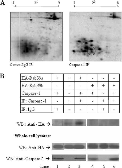
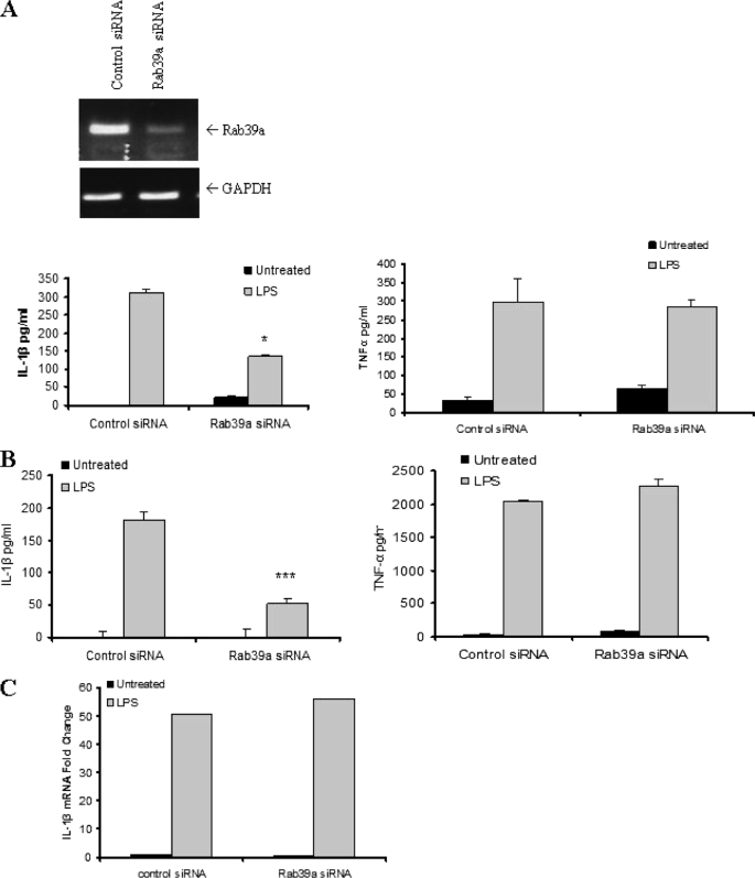
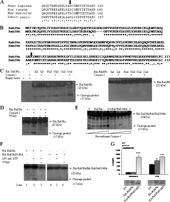
Interleukin-1beta (IL-1beta) is an important pro-inflammatory cytokine that is secreted by unconventional means in a caspase-1-dependent manner. Using a one-step immunoprecipitation approach to isolate endogenous caspase-1 from the monocytic THP1 cell line, we identified previously undescribed binding partners using mass spectrometry. One of the proteins identified was Rab39a, a member of the Rab GTPase family, a group of proteins that have important roles in protein trafficking and secretion. We confirmed by co-immunoprecipitation that Rab39a binds caspase-1. Knock down of Rab39a with small interfering RNA resulted in diminished levels of secreted IL-1beta but had no effect on induction of pro-IL-1beta mRNA by lipopolysaccharide. Rab39a contains a highly conserved caspase-1 cleavage site and was cleaved in the presence of recombinant caspase-1 or lipopolysaccharide. Finally, overexpression of Rab39a results in an increase in IL-1beta secretion, and furthermore, overexpression of a Rab39a construct lacking the caspase-1 cleavage site leads to an additional increase in IL-1beta secretion. Altogether, our findings show that Rab39a interacts with caspase-1 and suggest that Rab39a functions as a trafficking adaptor linking caspase-1 to IL-1beta secretion.


Figures

FIGURE 1.
FIGURE 1.
Rab39a is a novel binding partner of caspase-1. A, caspase-1 complexes were isolated from THP1 cell-free extracts. Proteins co-precipitating (IP) with caspase-1 were compared with proteins co-precipitating from an IgG control and were analyzed by two-dimensional gel electrophoresis (pI 5–8 first dimension and 12% SDS-PAGE second dimension). Protein spots of interest were identified by mass spectrometry. Rab39a was identified in the indicated area. B, 293T cells were transiently transfected with plasmids encoding caspase-1 and HA-Rab39a or HA-Rab39b. Caspase-1 was immunoprecipitated, and samples were probed with HA to show interaction. Whole cell lysates were immunoblotted (WB) with anti-HA anti-caspase-1. Results shown are each representative of two to four experiments.
FIGURE 2.
FIGURE 2.
Rab39a is required for IL-1β secretion. A, knock down of Rab39a is shown by PCR (upper panel). THP1 cells were transfected with siRNA to Rab39a for 48 h. Cells were stimulated with 100 ng/ml LPS overnight before harvesting. IL-1β and TNF-α levels were measured by ELISA. B, PBMCs were transfected with siRNA to Rab39a for 48 h. Cells were stimulated with 100 ng/ml LPS overnight before harvesting. IL-1β and TNF-α levels were measured by ELISA. A and B, results are expressed as mean ± S.D. for triplicate determinations. ***, p < 0.005; *, p < 0.05. All results are representative of three separate experiments. C, RNA from THP1 cells from siRNA experiments was extracted, and mRNA levels of IL-1β were measured by real time PCR. Expression of IL-1β mRNA was normalized to that of glyceraldehyde-3-phosphate dehydrogenase (GAPDH) mRNA. Data were calculated as fold change and are presented relative to that of untreated controls. Results shown are each representative of at least three independent experiments.
FIGURE 3.
FIGURE 3.
Rab39a contains a putative caspase-1 cleavage site and can be cleaved by recombinant caspase-1. A, caspase-1 cleavage is highly conserved. The arrow indicates the conserved putative caspase-1 cleavage site (see expasy peptide cutter site on the web). B, ClustalW alignment of the amino acid sequence of Rab39a and Rab39b is shown. The predicted cleavage site is indicated by the arrow (see expasy peptide cutter site on the web). C, 293T cells were transfected with plasmids encoding HA-Rab39a or HA-Rab39b. Cells were lysed, and increasing volumes of lysate were incubated with recombinant caspase-1 at 30 °C for 90 min. D, 293T cells were transfected with HA-Rab39a in the presence or absence of 100 μm caspase-1 inhibitor (YVAD-Cmk) and then incubated with recombinant caspase-1 at 30 °C for 90 min. E, 293T cells were transfected with HA-Rab39a or HA-Rab39a D148A. Lysates were incubated at 30 °C for 90 min with increasing concentrations of recombinant caspase-1. F, Raw 264.7 cells were transfected with plasmids encoding HA-Rab39a or HA-Rab39aD148A and treated with 100 ng/ml LPS and 5 mm ATP in the presence or absence of 100 μm caspase-1 inhibitor (YVAD-Cmk). Rab39a was detected by immunoblotting for anti-HA. G, HA-Rab39a or HA-Rab39aD148 was transfected into THP1 cells for 48 h. Cells were stimulated with 100 ng/ml LPS overnight before harvesting. Results are expressed as mean ± S.D. for triplicate determinations. ***, p < 0.005; *, p < 0.05. Results are representative of two or three independent experiments. EV, empty vector.
FIGURE 4.
FIGURE 4.
Rab39a can be induced by pro-inflammatory stimuli. A, Rab39a promoter contains numerous putative transcription factor binding sites as indicated (adapted from the Genomatix website). B, THP1 cells were stimulated with 100 ng/ml LPS or 5 nm Malp-2 or 1 μg/ml Pam3Cys for the indicated times. mRNA levels were measured by real time PCR with primers specific for Rab39a and Rab39b; expression is normalized to that of glyceraldehyde-3-phosphate dehydrogenase and is presented relative to that of untreated controls. Results shown are representative of three independent experiments.

References

    1. Thornberry N. A., Bull H. G., Calaycay J. R., Chapman K. T., Howard A. D., Kostura M. J., Miller D. K., Molineaux S. M., Weidner J. R., Aunins J., Elliston K., Ayala J., Casano F., Chin J., Ding G., Egger L., Gaffney E., Limjuco W., Palyha O., Raju S., Rolando A., Salley J., Yamin T., Lee T., Shively J., MacCross M., Mumford R., Schmidt J., Tocci M. (1992) Nature 356, 768–774 - PubMed
    1. Eder C. (2009) Immunobiology 214, 543–553 - PubMed
    1. Andrei C., Dazzi C., Lotti L., Torrisi M. R., Chimini G., Rubartelli A. (1999) Mol. Biol. Cell 10, 1463–1475 - PMC - PubMed
    1. Andrei C., Margiocco P., Poggi A., Lotti L. V., Torrisi M. R., Rubartelli A. (2004) Proc. Natl. Acad. Sci. U.S.A. 101, 9745–9750 - PMC - PubMed
    1. MacKenzie A., Wilson H. L., Kiss-Toth E., Dower S. K., North R. A., Surprenant A. (2001) Immunity 15, 825–835 - PubMed

Publication types

MeSH terms

LinkOut - more resources