. 2009 Dec 4;284(49):33957-65.
doi: 10.1074/jbc.M109.056309. Epub 2009 Oct 15.

Translocation and endocytosis for cell-penetrating peptide internalization

Affiliations

Translocation and endocytosis for cell-penetrating peptide internalization

Chen-Yu Jiao et al. J Biol Chem. .

Abstract

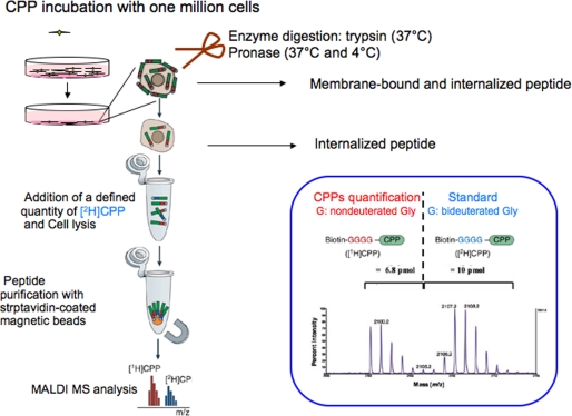
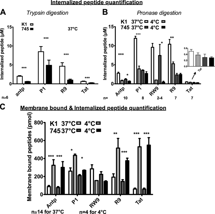
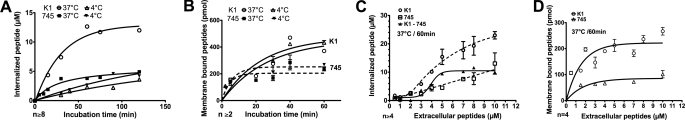
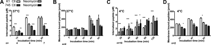
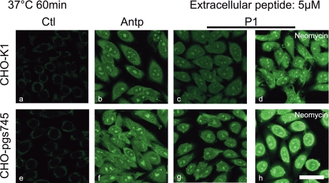
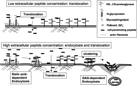
Cell-penetrating peptides (CPPs) share the property of cellular internalization. The question of how these peptides reach the cytoplasm of cells is still widely debated. Herein, we have used a mass spectrometry-based method that enables quantification of internalized and membrane-bound peptides. Internalization of the most used CPP was studied at 37 degrees C (endocytosis and translocation) and 4 degrees C (translocation) in wild type and proteoglycan-deficient Chinese hamster ovary cells. Both translocation and endocytosis are internalization pathways used by CPP. The choice of one pathway versus the other depends on the peptide sequence (not the number of positive changes), the extracellular peptide concentration, and the membrane components. There is no relationship between the high affinity of these peptides for the cell membrane and their internalization efficacy. Translocation occurs at low extracellular peptide concentration, whereas endocytosis, a saturable and cooperative phenomenon, is activated at higher concentrations. Translocation operates in a narrow time window, which implies a specific lipid/peptide co-import in cells.


Figures

FIGURE 1.
FIGURE 1.
Cell-penetrating peptide sequences used in this study. Biot, biotin.
FIGURE 2.
FIGURE 2.
Protocol of peptide quantification. The protocol used in this study is described under “Experimental Procedures.” Further detail can be found in Refs. and .
FIGURE 3.
FIGURE 3.
Quantification of internalized and membrane-bound cell penetrating peptides in CHO-K1 and CHO-pgsA-745 cells. A, internalized concentrations in wild type (K1) and xylosyltransferase I-deficient (745) CHO cells. One million cells were incubated with peptides for 75 min at 37 °C. The membrane-bound peptide was digested with trypsin. B, temperature effect on the amount of internalized peptide. One million cells were incubated with peptides during 75 min at 37 or 4 °C. The membrane-bound peptide was digested with pronase. C, temperature effect on the membrane-bound and internalized peptides. One million cells were incubated with peptides during 75 min at 37 or 4 °C. Cells were washed three times with culture medium. Extracellular concentration of peptides in A, B, and C: 2 μm for (R/W)9; 5 μm for Antp, P1, and R9; and 7.5 μm for Tat. Columns from left to right, K1, 37 °C; K1, 4 °C; 745, 37 °C; and 745, 4 °C. Error bars are S.E. obtained from n independent experiments. Differences in peptide quantity measured at 37 and 4 °C were: *, significant (0.01 < p < 0.05); **, very significant (0.001 < p < 0.01); and ***, highly significant (p > 0.001).
FIGURE 4.
FIGURE 4.
Kinetics and concentration dependence. Kinetics of peptide internalization (A) and binding to membrane (B). CHO-K1 or CHO-pgsA-745 cells were incubated, at 37 or 4 °C, with 5 μm extracellular peptide (P1) for the indicated incubation times. Effect of extracellular peptide concentration on internalized (C) or membrane-bound (D) peptide quantity. CHO-K1 or CHO-pgsA-745 cells were incubated with different concentrations of peptide (P1) during 60 min at 37 or 4 °C. Error bars are S.E. obtained from n independent experiments.
FIGURE 5.
FIGURE 5.
Effect of neomycin on peptide internalization (A and C) or binding to membrane (B and D). CHO-K1 or CHO-pgsA-745 cells were pretreated with 10 mm neomycin for 30 min, washed, and additionally incubated at 37 °C (A and B) or 4 °C (C and D) with 5 μm extracellular peptide (P1) for the indicated incubation times. Error bars are S.E. obtained from n independent experiments.
FIGURE 6.
FIGURE 6.
Confocal microscopy of Antp and P1 internalization at 37 °C. CHO-K1, CHO-pgsA-745, plated on glass coverslips, were incubated for 60 min with Antp (b and f) or P1 (c, d, g, and h) (5 μm). For d and e, cells were preincubated with 10 mm neomycin for 30 min. Cells were fixed with paraformaldehyde; biotinylated peptides were labeled with streptavidin-Alexa Fluor 488. Scale bar, 50 μm. a, control cells without peptide incubation.
FIGURE 7.
FIGURE 7.
Schematic representation of the different pathways of Antp internalization in cells. At submicromolar concentrations, Antp translocates into cells. Translocation is accomplished by a co-transport mechanism, likely a negatively charged membrane lipid. At higher concentrations, in addition to translocation, the peptide accumulates on the cell surface, leading to GAG clustering and endocytosis.

References

    1. Derossi D., Joliot A. H., Chassaing G., Prochiantz A. (1994) J. Biol. Chem. 269, 10444–10450 - PubMed
    1. Duchardt F., Fotin-Mleczek M., Schwarz H., Fischer R., Brock R. (2007) Traffic 8, 848–866 - PubMed
    1. Richard J. P., Melikov K., Vives E., Ramos C., Verbeure B., Gait M. J., Chernomordik L. V., Lebleu B. (2003) J. Biol. Chem. 278, 585–590 - PubMed
    1. Vivès E., Schmidt J., Pèlegrin A. (2008) Biochim Biophys Acta 1786, 126–138 - PubMed
    1. Console S., Marty C., García-Echeverría C., Schwendener R., Ballmer-Hofer K. (2003) J. Biol. Chem. 278, 35109–35114 - PubMed

Publication types

LinkOut - more resources