. 2009 Dec 11;284(50):34839-48.
doi: 10.1074/jbc.M109.035683. Epub 2009 Oct 15.

Leukocyte Ig-like receptor B4 (LILRB4) is a potent inhibitor of FcgammaRI-mediated monocyte activation via dephosphorylation of multiple kinases

Affiliations

Leukocyte Ig-like receptor B4 (LILRB4) is a potent inhibitor of FcgammaRI-mediated monocyte activation via dephosphorylation of multiple kinases

Hao Kim Lu et al. J Biol Chem. .

Abstract

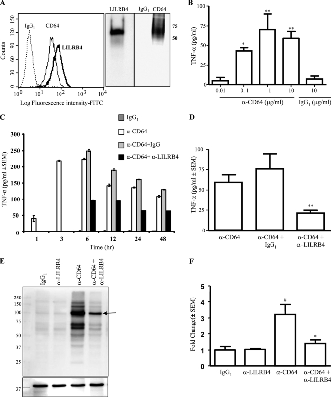
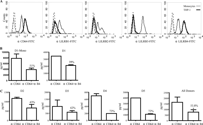
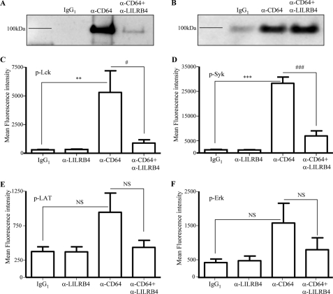
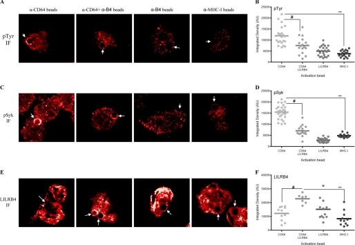
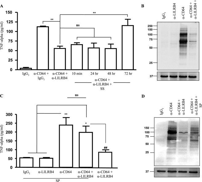
The leukocyte immunoglobulin-like receptor (LILR) B4 belongs to a family of cell surface receptors that possesses cytoplasmic immunoreceptor tyrosine-based inhibitory motifs (ITIMs). LILRB4 is believed to down-regulate activation signals mediated by non-receptor tyrosine kinase cascades through the recruitment of SHP-1. However, the exact mechanisms of LILRB4-mediated inhibition are not fully elucidated. In this study, we demonstrate high level surface expression of LILRB4 on THP-1 cells and primary peripheral blood monocytes, which profoundly inhibited production of a key pro-inflammatory cytokine (TNFalpha) induced by FcgammaRI (CD64). We also report that LILRB4 aggregated to sites of activation upon co-ligation with CD64 and that this may enhance its inhibitory effects. Cross-linking of CD64 on THP-1 cells markedly increased phosphorylation of multiple proteins including tyrosine kinases and signaling molecules (Lck, Syk, LAT, and Erk), an adaptor protein that targets protein-tyrosine kinases for degradation (c-Cbl) and a protein involved in the formation of actin cytoskeletal rearrangement (alpha-actinin-4). Co-ligation of LILRB4 considerably reduced CD64-mediated phosphorylation of Lck, Syk, LAT, Erk, and c-Cbl but not alpha-actinin-4, suggesting selective inhibition of signaling molecules. Treatment of cells with a broad-spectrum phosphatase inhibitor, sodium pervanadate (SP), significantly reversed LILRB4-mediated inhibition of TNFalpha production and protein tyrosine phosphorylation. In comparison, treatment with an SHP-1 specific inhibitor, sodium stibogluconate (SS) has no effects indicating involvement of phosphatase(s) other than SHP-1 in LILRB4 signaling. Collectively, our data show LILRB4 is a potent inhibitor of monocytes activation. This may provide a new potential therapeutic strategy for inflammatory conditions characterized by excessive TNFalpha production.


Figures

FIGURE 1.
FIGURE 1.
Co-ligation of LILRB4 reduced CD64-mediated TNFα production and protein-tyrosine phosphorylation on THP-1 cells. A, representative data showing comparable surface expression of LILRB4 and CD64, as determined by flow cytometry and confirmed by immunoprecipitation. B, TNFα ELISA on culture supernatants from THP-1 cells cross-linked with anti-CD64 monoclonal antibody for 24 h (n = 4); *, p < 0.05; **, p < 0.01 as compared with IgG1 control. C, TNFα ELISA on culture supernatants from THP-1 cells co-ligated with saturation amounts of anti-CD64 and anti-LILRB4 monoclonal antibodies (10 μg/ml each) at various time points. D, shows a summary of four independent TNFα ELISA experiments at the 24-h time point; **, p < 0.01 compared with cells treated with anti-CD64 alone. E, representative anti-phosphotyrosine (anti-pTyr) Western blot of THP-1 cell lysates after a 2 min cross-linking with control IgG1 (lane 1), anti-LILRB4 (lane 2), anti-CD64 (lane 3), or a combination of anti-CD64 and anti-LILRB4 (lane 4) monoclonal antibodies (10 μg/ml each). The lower panel shows the same membrane blotted for β-actin. F, densitometry of three independent anti-pTyr Western blots; #, p < 0.05 compared with IgG1 control; *, p < 0.05 anti-CD64 versus anti-CD64+anti-LILRB4.
FIGURE 2.
FIGURE 2.
Co-ligation of LILRB4 and CD64 on magnetic bead purified or non-purified peripheral blood monocytes significantly abrogated TNFα production. A, representative data on the expression of CD64 and the inhibitory LILRs B1, B2, B3, and B4 on THP-1 cells and primary monocytes as determined by flow cytometry (n = 5). Both THP-1 cells and primary monocytes expressed comparable levels of CD64. However, unlike primary monocytes that expressed all inhibitory LILRs, THP-1 cells selectively expressed high levels of LILRB4. The dotted histograms on the left are corresponding isotype-matched negative controls. B, TNFα ELISA of culture supernatants from 5 × 10⁴ magnetic beads separated monocytes or corresponding peripheral blood mononuclear cells that contained 5 × 10⁴ monocytes after co-ligation of CD64 and LILRB4 for 24 h. C, TNFα ELISA of culture supernatants from peripheral blood mononuclear cells (5 × 10⁴ monocytes) from four independent donors that were activated after co-ligation of CD64 and LILRB4 for 24 h. Note that there are substantial differences among individuals in the amounts of TNFα produced in response to CD64 cross-linking. *, p < 0.05 as compared with cells treated with anti-CD64 alone.
FIGURE 3.
FIGURE 3.
CD64-mediated phosphorylation of c-Cbl and multiple protein-tyrosine kinases but not α-actinin-4 is down-regulated by LILRB4. A and B, THP-1 cells (5 × 10⁷) were lysed after 2 min of cross-linking with control IgG1 (lane 2), anti-CD64 (lane 3), or a combination of anti-CD64 and anti-LILRB4 (lane 4) monoclonal antibodies (10 μg/ml), followed by anti-pTyr immunoprecipitation. Immunoprecipitated proteins were Western blotted with anti-c-Cbl (A) or anti- α-actinin-4 (B) specific antibodies. C–F, Lck (C), Syk (D), LAT (E), and Erk (F) phosphotyrosine multiplex assay on THP-1 cell lysates after a 2-min cross-linking with same amounts of control IgG1, anti-LILRB4, anti-CD64 or a combination of anti-CD64 and anti-LILRB4 monoclonal antibodies. Error bars indicate S.E. of three independent experiments; **, p < 0.01; ***, p < 0.001; #, p < 0.05; ###, p < 0.001.
FIGURE 4.
FIGURE 4.
Phosphorylated proteins are increased at sites of receptor activation; LILRB4 is preferentially aggregated at sites of CD64 and LILRB4 co-ligation and down-regulates tyrosine phosphorylation. A, C, and E, representative immuno-fluorescence staining with anti-pTyr (A), anti-pSyk (C), or anti-LILRB4 (E) antibodies on cells treated with anti-CD64, anti-CD64+anti-LILRB4, anti-LILRB4 or control anti-MHC-class I-coated beads. Arrows indicate bead-receptor contact sites. B, D, and F, integrated density of pTyr (B), pSyk (D), and LILRB4 (E) at bead-receptor contact sites. Each dot represents a contact site on an individual cell, and mean values are indicated by horizontal lines. **, p < 0.01; #, p < 0.05.
FIGURE 5.
FIGURE 5.
LILRB4 signaling is differentially regulated by SS and SP. A, TNFα ELISA on culture supernatants from THP-1 cells cross-linked for 6 h using control IgG1, anti-CD64+IgG1, or anti-CD64+anti-LILRB4 after pretreatment with or without 11 μm SS for 10 min, 24 h, 48 h, or 72 h (n = 3) (**, p < 0.01). B, a representative anti-pTyr Western blot on cell lysates from THP-1 cells that were pretreated with 11 μm SS for 72 h and cross-linked for 2 min with the indicated antibodies. C, TNFα ELISA on culture supernatants from THP-1 cells cross-linked for 6 h using control IgG1, anti-CD64+IgG1, or anti-CD64+anti-LILRB4 after pretreatment with 100 μm SP for 10 min (n = 4) (*, p < 0.05; **, p < 0.01; ##, p < 0.01). D, representative anti-pTyr Western blot on cell lysates from THP-1 cells that were pretreated with or without 100 μm of SP for 10 min and cross-linked for 2 min with the indicated antibodies. The bottom panels in C and D show the same membranes blotted for β-actin to ensure equal protein loading.

Similar articles

Cited by

References

    1. Hamerman J. A., Lanier L. L. (2006) Sci. STKE 320, re1. - PubMed
    1. Cambier J. C. (1995) Immunol. Today 16, 110. - PubMed
    1. Bu J. Y., Shaw A. S., Chan A. C. (1995) Proc. Natl. Acad. Sci. U.S.A. 92, 5106–5110 - PMC - PubMed
    1. Billadeau D. D., Leibson P. J. (2002) J. Clin. Invest. 109, 161–168 - PMC - PubMed
    1. Chacko G. W., Brandt J. T., Coggeshall K. M., Anderson C. L. (1996) J. Biol. Chem. 271, 10775–10781 - PubMed