. 2009 Dec;297(6):G1041-52.
doi: 10.1152/ajpgi.00328.2009. Epub 2009 Oct 15.

Oral PEG 15-20 protects the intestine against radiation: role of lipid rafts

Affiliations

Oral PEG 15-20 protects the intestine against radiation: role of lipid rafts

Vesta Valuckaite et al. Am J Physiol Gastrointest Liver Physiol. 2009 Dec.

Abstract

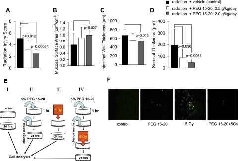
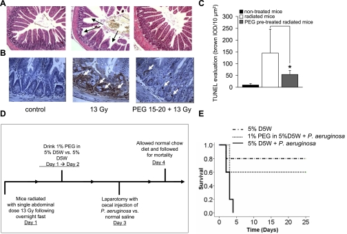
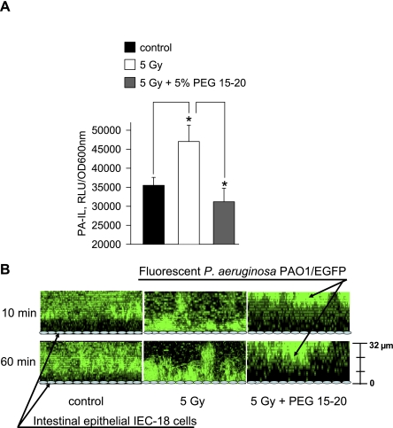
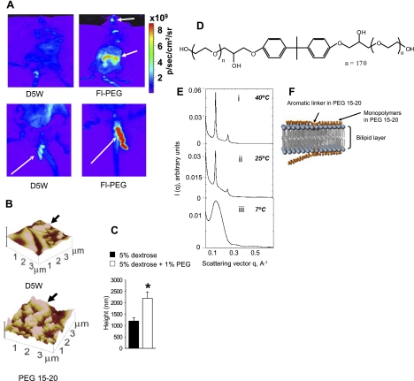
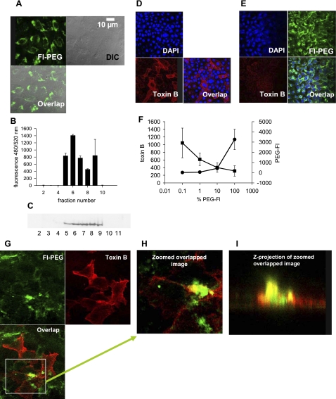
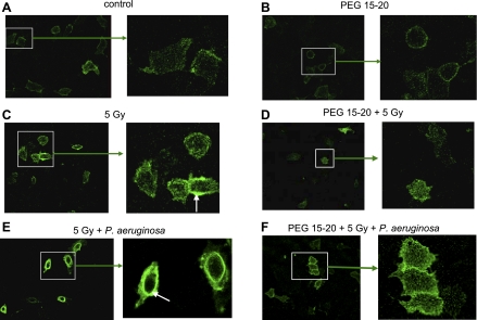
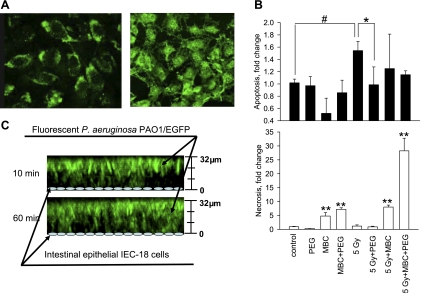
Intestinal injury following abdominal radiation therapy or accidental exposure remains a significant clinical problem that can result in varying degrees of mucosal destruction such as ulceration, vascular sclerosis, intestinal wall fibrosis, loss of barrier function, and even lethal gut-derived sepsis. We determined the ability of a high-molecular-weight polyethylene glycol-based copolymer, PEG 15-20, to protect the intestine against the early and late effects of radiation in mice and rats and to determine its mechanism of action by examining cultured rat intestinal epithelia. Rats were exposed to fractionated radiation in an established model of intestinal injury, whereby an intestinal segment is surgically placed into the scrotum and radiated daily. Radiation injury score was decreased in a dose-dependent manner in rats gavaged with 0.5 or 2.0 g/kg per day of PEG 15-20 (n = 9-13/group, P < 0.005). Complementary studies were performed in a novel mouse model of abdominal radiation followed by intestinal inoculation with Pseudomonas aeruginosa (P. aeruginosa), a common pathogen that causes lethal gut-derived sepsis following radiation. Mice mortality was decreased by 40% in mice drinking 1% PEG 15-20 (n = 10/group, P < 0.001). Parallel studies were performed in cultured rat intestinal epithelial cells treated with PEG 15-20 before radiation. Results demonstrated that PEG 15-20 prevented radiation-induced intestinal injury in rats, prevented apoptosis and lethal sepsis attributable to P. aeruginosa in mice, and protected cultured intestinal epithelial cells from apoptosis and microbial adherence and possible invasion. PEG 15-20 appeared to exert its protective effect via its binding to lipid rafts by preventing their coalescence, a hallmark feature in intestinal epithelial cells exposed to radiation.


Figures

Fig. 1.
Fig. 1.
High-molecular-weight polyethylene glycol 15,000–20,000 Da (PEG 15–20) protects the rat intestine and cultured rat intestinal epithelial cells (IECs) against radiation exposure. AD: dose-dependent protective effect of oral PEG 15–20 on the rat intestine within the scrotum exposed to daily fractionated radiation. Data are means ± SD; n = 9–12. E: experimental design for cultured rat IECs (IEC-18) exposed to radiation. I: control IEC-18 cells, no treatment. II: PEG-treated cells, IEC-18 cells are exposed to 5% PEG 15–20 for 1 h, followed by gentle washing with culture media, and analyzed in 24 h. III: 5 Gy, IEC-18 cells are exposed to 5 Gy radiation; analysis performed in 24 h. IV: PEG 15–20 + 5 Gy, IEC-18 cells are exposed to 5% PEG 15–20 for 1 h, followed by gentle washing with culture media, and exposed to 5 Gy radiation; analysis performed in 24 h. F: nuclear DNA fragmentation detected by TUNEL assay in IEC-18 cells at 24 h posttreatment.
Fig. 2.
Fig. 2.
Mice drinking oral PEG 15–20 are protected against radiation-induced apoptosis and display enhanced resistance to postradiation infection. A: Hematoxylin and eosin staining of ileum cross sections demonstrates morphological changes in the intestinal epithelium following abdominal irradiation (13 Gy) with a decrease in the number and height of villous projections (villous blunting, shown by black arrows) that are prevented when mice drink 1% PEG 15–20 (PEG 15–20 + 13 Gy). B: TUNEL assay of intestinal epithelial cross sections. White arrows are focused on apoptotic cells. C: quantitative assessment of apoptosis performed by measurement of the brown stained area per 10 μm2 using Automated Cellular Imaging Software, n = 90 images from 3 mice/group, *P < 0.001. D: experimental design of postradiation infection. E: Kaplan-Mayer survival curves demonstrating enhanced resistance against postradiation intestinal exposure to Pseudomonas aeruginosa (P. aeruginosa) when mice drink oral PEG 15–20 (n = 10/group, P < 0.001). D5W, 5% dextrose solution in water. IOD, integrated optical density.
Fig. 3.
Fig. 3.
PEG 15–20 prevents P. aeruginosa adherence to and possible invasion of IEC-18 cells and suppresses virulence expression in P. aeruginosa when exposed to media from radiated IEC-18 cells. A: expression of PA-I lectin/adhesin in P. aeruginosa PAO1/lecA::lux exposed to media collected from IEC-18 cells following various treatments. Data represent luminescence values normalized to bacterial cell density (OD 600 nm). PA-I lectin expression is significantly increased in P. aeruginosa when exposed to media from irradiated compared with nonradiated cells and significantly decreased when exposed to media from irradiated cells pretreated with PEG 15–20. Data are means ± SD; n = 4, *P < 0.005. B: z-plane reconstructions of multiple stacked images of P. aeruginosa PAO1/enhanced green fluorescence protein (EGFP) collected at 10 and 60 min, demonstrating spatial orientation of P. aeruginosa to the IEC-18 cell surface. Green fluorescence represents fluorescent P. aeruginosa cells. The adhesion/invasion of P. aeruginosa to radiated IEC-18 cells is abundant whereby bacteria can be seen suspended above and repelled away from the IEC-18 cell surface when cells were exposed to PEG 15–20 followed by washing with new media. RLU, relative light units.
Fig. 4.
Fig. 4.
PEG 15–20 coats the luminal surface of the gastrointestinal tract and associates within bilipid membranes. A: Xenogen IVIS 200 images of live mice drinking D5W or 1% fluorescein-labeled PEG 15–20 in D5W (Fl-PEG). Top: images of whole mice, arrows on fluorescent spots demonstrating the distribution of the polymer in mouth (pharynx) and midgut/hindgut. Bottom: Xenogen images of expelled feces. B: atomic force microscopy (AFM) images of midgut segments of intestine of mice drinking D5W (top) or 1% PEG 15–20 in D5W (bottom). C: AFM height deflection measurements; n = 3, *P < 0.05. D: structure of PEG 15–20. E: Synchrotron small-angle X-ray scattering profiles collected at three temperatures, 40°C, 25°C, and 7°C, on samples prepared with PEG 15–20. The quaternary composition consisted of 0.748 weight fraction water, Φw; 0.0961 weight fraction dimyristoyl-sn-glycero-3-phosphocholine, ΦL; 0.1333 weight fraction of polymer, Φp; and 0.0221 weight fraction lauryldimethylamine-N-oxide surfactant, Φs. q, scattering vector; I(q), the intensity as a function of the magnitude q of the scattering vector. F: schematic illustration of PEG 15–20 association with model biological membrane.
Fig. 5.
Fig. 5.
PEG binds to lipid rafts and competes for binding to lipid rafts with the Cholera toxin B. A: image of the IEC-18 cell coincubated with Fl-PEG. Top, left: fluorescent confocal image. Top, right: differential interference contrast (DIC) image. Bottom: merged image. B: fluorescence measured in sucrose gradient fractions of IEC-18 cells coincubated for 1 h with Fl-PEG followed by vigorous washing. C: Western blot analysis of sucrose gradient fractions isolated from IEC-18 cells pretreated for 1 h with the Fl-PEG. Purified mouse anti-Flotillin 1 MAb (BD Transduction Laboratories, San Diego, CA) were used in the analysis. D: fluorescent confocal images of IEC-18 cells stained with Cholera toxin B to identify lipid rafts. Top, left: nuclear DAPI staining. Bottom, left: lipid raft staining using red fluorescent protein-fused Cholera toxin B. Bottom, right: merged image. E: fluorescent confocal images of IEC-18 cells pretreated for 1 h with 0.25% Fl-PEG followed by coincubation with red fluorescent protein fused Cholera toxin B. Top, left: nuclear DAPI staining. Top, right: green fluorescence image. Bottom, left: red fluorescence image. Bottom, right: merged image. F: dose-dependent effect of Fl-PEG on Cholera toxin B binding to lipid rafts. Red fluorescence (toxin B, ■) or green fluorescence (PEG-Fl, •) intensity were assessed using the Image J program. Data are means ± SD, n = 10. G: fluorescent confocal images of IEC-18 cells pretreated for 1 h with 0.2% Fl-PEG and stained with Cholera toxin B for the lipid rafts. Top, left: green fluorescence image. Bottom, left: red fluorescence image. Top, right: merged image. H: enlarged image of green/red colocalization from G. I: z-plane image of selected area from H.
Fig. 6.
Fig. 6.
PEG 15–20 prevents the coalescence of lipid rafts in IEC-18 cells. A: control IEC-18 cells. B: IEC cells exposed to PEG 15–20 for 1 h. C: IEC-18 cells exposed to 5 Gy irradiation. D: IEC-18 cells pretreated with PEG 15–20, followed by 5 Gy irradiation. E: IEC-18 cells exposed to 5 Gy irradiation followed by infection with P. aeruginosa PAO1 for 1 h. F: IEC-18 cells pretreated with PEG 15–20, followed by 5 Gy irradiation and infection with P. aeruginosa PAO1. White arrows indicate lipid raft coalescence.
Fig. 7.
Fig. 7.
Methyl-B-cyclodextrin (MBC) treatment of IEC-18 cells abrogates the protective effect of PEG 15–20. A: images of IEC-18 cells coincubated with 0.25% Fl-PEG without (left) and with (right) MBC. B: DNA fragmentation (cell death ELISA) measured in IEC-18 cells (apoptosis) and IEC-18 conditioned media (necrosis) (n = 5/group, *P < 0.005, **P < 0.001). C: z-plane reconstructions of multiple stacked images of P. aeruginosa PAO1/EGFP demonstrating spatial orientation of P. aeruginosa to MBC-treated radiated IEC-18 cells. Images demonstrate that depletion of cholesterol attenuates the protective effect of PEG in its ability to repel bacteria away from the intestinal epithelial surface. At 10 min, scattered bacteria are seen attached to IEC-18 cells, which is observed to be more prominent at 60 min.

Similar articles

Cited by

References

    1. Baddeley AJ, Gundersen HJ, Cruz-Orive LM. Estimation of surface area from vertical sections. J Microsc 142: 259–276, 1986 - PubMed
    1. Bertz H, Auner HW, Weissinger F, Salwender HJ, Einsele H, Egerer G, Sandherr M, Schuttrumpf S, Sudhoff T, Maschmeyer G. Antimicrobial therapy of febrile complications after high-dose chemo-/radiotherapy and autologous hematopoietic stem cell transplantation—guidelines of the Infectious Diseases Working Party (AGIHO) of the German Society of Hematology and Oncology (DGHO). Ann Hematol 82, Suppl 2: S167–S174, 2003 - PubMed
    1. Bionda C, Hadchity E, Alphonse G, Chapet O, Rousson R, Rodriguez-Lafrasse C, Ardail D. Radioresistance of human carcinoma cells is correlated to a defect in raft membrane clustering. Free Radic Biol Med 43: 681–694, 2007 - PubMed
    1. Brook I, Elliott TB, Ledney GD, Knudson GB. Management of postirradiation sepsis. Mil Med 167: 105–106, 2002 - PubMed
    1. Brook I, Elliott TB, Ledney GD, Shoemaker MO, Knudson GB. Management of postirradiation infection: lessons learned from animal models. Mil Med 169: 194–197, 2004 - PubMed

Publication types

MeSH terms