. 2009 Nov 3;106(44):18809-14.
doi: 10.1073/pnas.0908767106. Epub 2009 Oct 15.

TDP-43 mutant transgenic mice develop features of ALS and frontotemporal lobar degeneration

Affiliations

TDP-43 mutant transgenic mice develop features of ALS and frontotemporal lobar degeneration

Iga Wegorzewska et al. Proc Natl Acad Sci U S A. .

Abstract

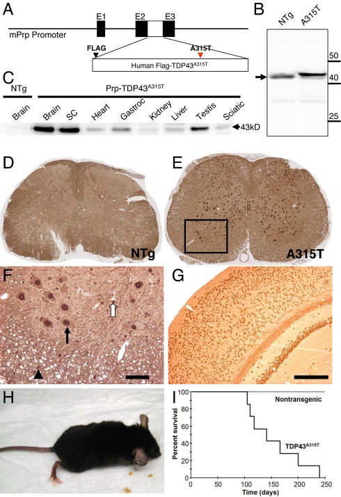
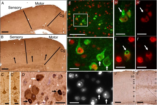
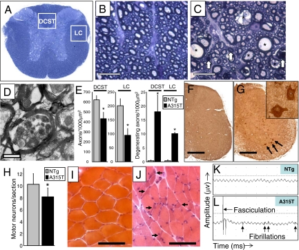
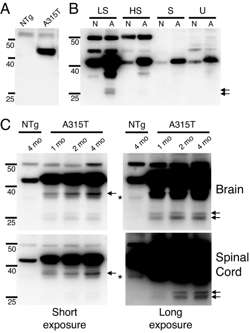
Frontotemporal lobar degeneration (FTLD) and amyotrophic lateral sclerosis (ALS) are neurodegenerative diseases that show considerable clinical and pathologic overlap, with no effective treatments available. Mutations in the RNA binding protein TDP-43 were recently identified in patients with familial amyotrophic lateral sclerosis (ALS), and TDP-43 aggregates are found in both ALS and FTLD-U (FTLD with ubiquitin aggregates), suggesting a common underlying mechanism. We report that mice expressing a mutant form of human TDP-43 develop a progressive and fatal neurodegenerative disease reminiscent of both ALS and FTLD-U. Despite universal transgene expression throughout the nervous system, pathologic aggregates of ubiquitinated proteins accumulate only in specific neuronal populations, including layer 5 pyramidal neurons in frontal cortex, as well as spinal motor neurons, recapitulating the phenomenon of selective vulnerability seen in patients with FTLD-U and ALS. Surprisingly, cytoplasmic TDP-43 aggregates are not present, and hence are not required for TDP-43-induced neurodegeneration. These results indicate that the cellular and molecular substrates for selective vulnerability in FTLD-U and ALS are shared between mice and humans, and suggest that altered DNA/RNA-binding protein function, rather than toxic aggregation, is central to TDP-43-related neurodegeneration.


Conflict of interest statement

The authors declare no conflict of interest.

Figures

Fig. 1.
Fig. 1.
Expression of a disease mutant form of TDP-43 throughout the nervous system in mice leads to progressive gait disturbance, and premature death. (A) Schematic diagram of the Prp-TDP43A315T construct. A cDNA encoding a Flag-tagged human TDP-43 protein with the A315T point mutation seen in patients with familial ALS was placed into the mouse Prion protein promoter construct (mPrp). (B) Western blotting of brain lysates from nontransgenic (NTg) and Prp-TDP43A315T mice (A315T) using an anti-TDP43 antibody showed a slight shift to a higher molecular weight due to the presence of the Flag tag, and approximately 3-fold higher levels compared to endogenous TDP-43 (arrow). (C) Western blotting of tissue lysates with anti-Flag antibody (specific for the human transgene) showed highest levels in brain, spinal cord, and testis, with lower levels in skeletal muscle (Gastroc–gastrocnemius), heart and other tissues. (D–G) Immunohistochemistry with anti-Flag antibody showed expression of the TDP43A315T protein throughout the spinal cord (E–“A315T”), which was absent from nontransgenic animals (D–“NTg”). (F) Higher power image of area corresponding to the boxed region in E, showing that TDP43A315T mutant protein was present in nuclei of large ventral horn motor neurons (arrow), as well as smaller nuclei in white matter tracts (arrowhead) and throughout the neuropil (open arrow) presumably corresponding to oligodendroglia and astrocytes. (Scale bar, 50 μm.) (G) Similar widespread expression was observed in the brain in neurons and non-neuronal cells, including all layers of cortex and hippocampus. (Scale bar, 200 μm.) (H) Photograph of end-stage Prp-TDP43A315T mouse (≈5 months old), unable to move its hindlimbs or right itself when placed on its back. (I) Survival curve of Prp-TDP43A315T mice showed an average survival of 153 days. No Prp-TDP43A315T mice have survived beyond 240 days.
Fig. 2.
Fig. 2.
Selective brain pathology in Prp-TDP43A315T mice reminiscent of FTLD-U. (A–D) Ubiquitin immunohistochemical analysis of a non-transgenic (A) and late stage (4.5-month-old) Prp-TDP43A315T brain (B) showing ubiquitin pathology (arrows) selectively in a population of layer 5 neurons in motor, sensory and cingulate (Cg) cortex, despite universal expression of the transgene in all cortical layers and throughout the brain. (Scale bars, 300 μm.) (C and C′) Ubiquitin accumulation was predominantly in pyramidal neurons, as either discrete cytoplasmic aggregates (C) or diffusely increased cytoplasmic staining (C′). (Scale bars, 10 μm.) (D) Some neurons contained large organized ubiquitin-positive cytoplasmic inclusions (black arrows), whereas others contained multiple smaller aggregates (white arrow). (Scale bar, 10 μm.) (E–I) Double immunostaining with antibodies to the C terminus of TDP-43 (red) and ubiquitin (green). (E) Some neurons with ubiquitin pathology showed loss of nuclear TDP-43 staining. (Scale bar, 50 μm.) (F) Higher power image of boxed region from (E), showing neurons with diminished nuclear TDP-43 staining (arrows). (G) DAPI staining of same field as (F) showing that nuclear structure is intact in neurons that have lost TDP-43 staining (white arrows indicate the same cells in F and G), indicating loss of TDP-43 occurs before neuron degeneration. (H and I) Overlay image of double immunofluorescence for ubiquitin (green) and TDP-43 (red) showing typical large cytoplasmic ubiquitinated aggregates (white arrows). (H′ and I′) The same images showing only TDP-43 staining. Notably, ubiquitin-positive aggregates did not contain TDP-43. (Scale bars for F through I, 10 μm.) (J and J′) GFAP immunostaining was also selectively increased in cortical layer 5 of Prp-TDP43A315T mice (J′) as compared to controls (J), indicating a reactive astrocytosis in the region of degenerating neurons with ubiquitin pathology. (Scale bars, 200 μm.)
Fig. 3.
Fig. 3.
Prp-TDP43A315T mice develop motor neuron disease. (A) Toluidine blue stained section of lower thoracic spinal cord, indicating regions of descending motor pathways in the mouse, including the dorsal corticospinal tract (DCST) and lateral columns (LC). (B and C) Lateral columns from an end-stage nontransgenic Prp-TDP43A315T mouse (C) showed both large dilated axons (asterisks) and degenerating axons (white arrows) in descending motor axons in Prp-TDP43A315T mice that were not present in age-matched nontransgenic controls (B). (Scale bar, 10 μm.) (D) Electron micrograph of dorsal corticospinal tract axons from Prp-TDP43A315T mouse, showing accumulations of dark axoplasmic material in a degenerating corticospinal tract axon. (Scale bar, 500 nm.) (E) Fewer axons were present in the DCST and LC of Prp-TDP43A315T mice (A315T) as compared to nontransgenic controls (NTg), with more degenerating fibers seen in Prp-TDP43A315T mice (* = t test, P < 0.05). (F and G) Ubiquitin immunohistochemistry of spinal cord from a nontransgenic control (F) and a Prp-TDP43A315T mouse (G), showing an increase in ubiquitin staining in lower motor neurons (black arrows, and inset), as well as scattered interneurons in Prp-TDP43A315T mice. (Scale bars, 400 μm.) (H) Quantitation of motor neurons from the L3–L5 region showed an approximate 20% reduction in motor neurons in Prp-TDP43A315T mice compared to nontransgenic controls (* = t test, P < 0.01). Hematoxylin and eosin stain of muscle from a non-transgenic (I) and Prp-TDP43A315T mouse (J), showing scattered and grouped muscle fiber atrophy, characteristic of motor axon degeneration. (Scale bars, 60 μm.) Electromyography from a 4-month-old Prp-TDP43A315T mouse (L–A315T) showing fibrillations and fasciculations, common findings in ALS indicative of muscle fiber denervation and motor unit degeneration/regeneration. These findings were never present in nontransgenic (K–NTg) mice.
Fig. 4.
Fig. 4.
Prp-TDP43A315T mice show C-terminal fragmentation of TDP-43 in the presymptomatic phase. (A) Immunoblot of spinal cord lysates from 2-month-old non-transgenic (NTg) and Prp-TDP43A315T (A315T) mice using an antibody to the Flag epitope located at the N terminus of the transgene. Only a single band is observed at approximately 43 kDa. (B) TDP43 immunoblot of serial fractions of brain from a non-transgenic (N) and Prp-TDP43A315T mouse (A). Fractions shown are low salt (LS), high salt with Triton-X100 (HS), sarkosyl buffer (S), and urea buffer (U). Both full-length and C-terminal fragments (arrows) are predominant in the LS soluble fraction. (C) Immunoblots of the detergent soluble phase of brain and spinal cord lysates from either presymptomatic (1 month, 2 months) or late symptomatic (4 months) Prp-TDP43A315T mice, using a polyclonal anti-TDP43 antibody raised to amino acids 1–260 (of 414). On shorter exposures (left panels) multiple smaller fragments of TDP43 at approximately 35 kDa were seen (arrow), which were already present by 1 month of age in both brain and spinal cord. Longer exposures (right panel) also revealed two bands at approximately 24 kDa and 26 kDa (arrows) which were present before symptom onset, and appeared to increase during the disease course. A faint single band at approximately 34 kDa is seen in the age-matched nontransgenic control tissue on long exposure, which likely represents a known splice form of endogenous mouse TDP-43 (asterisk). However the 35 kDa, 26 kDa, and 24 kDa C-terminal fragments were never seen in non-transgenic littermate controls.

References

    1. Seeley WW. Selective functional, regional, and neuronal vulnerability in frontotemporal dementia. Curr Opin Neurol. 2008;21:701–707. - PMC - PubMed
    1. Cleveland DW, Rothstein JD. From Charcot to Lou Gehrig: Deciphering selective motor neuron death in ALS. Nat Rev Neurosci. 2001;2:806–819. - PubMed
    1. Pasinelli P, Brown RH. Molecular biology of amyotrophic lateral sclerosis: Insights from genetics. Nat Rev Neurosci. 2006;7:710–723. - PubMed
    1. Murphy JM, et al. Continuum of frontal lobe impairment in amyotrophic lateral sclerosis. Arch Neurol. 2007;64:530–534. - PubMed
    1. Lomen-Hoerth C, Anderson T, Miller B. The overlap of amyotrophic lateral sclerosis and frontotemporal dementia. Neurology. 2002;59:1077–1079. - PubMed

Publication types

MeSH terms

Substances