. 2009 Nov 3;106(44):18491-6.
doi: 10.1073/pnas.0907950106. Epub 2009 Oct 15.

Computational design of ligand binding is not a solved problem

Affiliations

Computational design of ligand binding is not a solved problem

Bettina Schreier et al. Proc Natl Acad Sci U S A. .

Abstract

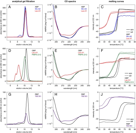
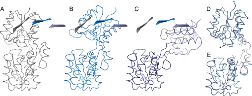
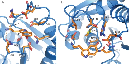
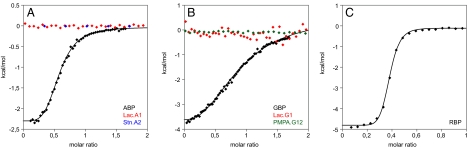
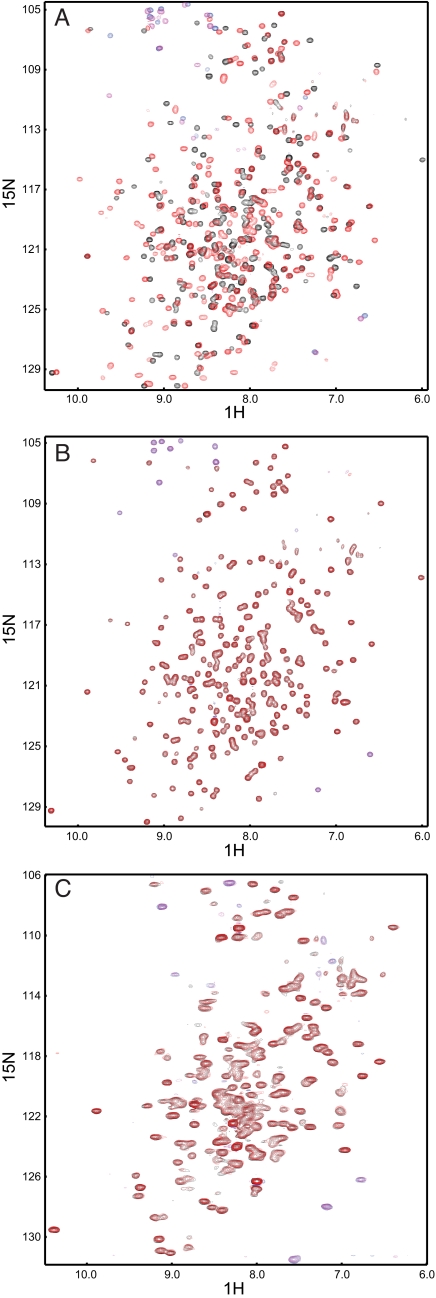
Computational design has been very successful in recent years: multiple novel ligand binding proteins as well as enzymes have been reported. We wanted to know in molecular detail how precise the predictions of the interactions of protein and ligands are. Therefore, we performed a structural analysis of a number of published receptors designed onto the periplasmic binding protein scaffold that were reported to bind to the new ligands with nano- to micromolar affinities. It turned out that most of these designed proteins are not suitable for structural studies due to instability and aggregation. However, we were able to solve the crystal structure of an arabinose binding protein designed to bind serotonin to 2.2 A resolution. While crystallized in the presence of an excess of serotonin, the protein is in an open conformation with no serotonin bound, although the side-chain conformations in the empty binding pocket are very similar to the conformations predicted. During subsequent characterization using isothermal titration calorimetry, CD, and NMR spectroscopy, no indication of binding could be detected for any of the tested designed receptors, whereas wild-type proteins bound their ligands as expected. We conclude that although the computational prediction of side-chain conformations appears to be working, it does not necessarily confer binding as expected. Hence, the computational design of ligand binding is not a solved problem and needs to be revisited.


Conflict of interest statement

The authors declare no conflict of interest.

Figures

Fig. 1.
Fig. 1.
Biophysical characteristics of designed receptor proteins: (A–C) ABP, Lac.A1, and Stn.A2, (D–F) GBP, Lac.G1, and PMPA.G12, (G–J) RBP and TNT.R1. (A) Association states measured by analytical gel filtration (0.5 mg loaded each). All three variants elute at the same volume, which corresponds to the molecular mass of monomeric protein. (B) CD spectra: the intensity of the signal of Lac.A1 is slightly reduced compared to ABP and Stn.A2. (C) Melting curves: no clear increase in stability upon addition of the respective ligand was observed for all three proteins. Thermal stability of Lac.A1 is severely reduced compared to wild-type, whereas the stability of Stn.A2 is almost the same. However, visible aggregation of Stn.A2 occurred, leading to varying signal intensity after unfolding. (D) Analytical gel filtration (0.6 mg each). Lac.G1 exists in solution only in the form of higher order oligomers. PMPA.G12 elutes in a single defined peak but with a higher apparent molecular weight than wild-type GBP. (E) CD spectra: a loss of secondary structure is observed in Lac.G1 compared to GBP and PMPA.G12. (F) Melting curves: A stability increase upon addition of ligand was only observed for wild-type GBP. No cooperative unfolding could be observed for Lac.G1. (G) Analytical gel filtration: (0.9 mg each). Monomeric TNT.R1 can be purified but aggregation increases over time. (H) CD spectra: TNT.R1 has a lower secondary structure content compared to RBP. (J) Melting curves: The thermal stability of TNT.R1 is severely reduced compared to wild type. Addition of TNT further decreases thermal stability, because of the solvent acetonitrile.
Fig. 2.
Fig. 2.
Open conformations of Stn.A2 compared to wild-type ABP (5abp). (A) ABP (gray ribbon) in closed conformation. (B) Stn.A2 molecule A (light blue ribbon). (C) Stn.A2 molecule B (dark blue ribbon). All structures are aligned over their C-terminal domains. One β-strand of the N-terminal domain is highlighted as cartoon for each structure and shown in each figure to illustrate the hinge bending motion. (D) Superimposition of the N-terminal domains of ABP, and Stn.A2 molecule A and B. Loop 12–19 (includes design residues A16 and S17) adopts a different conformation in Stn.A2 molecule B (indicated by an asterisk). (E) Superimposition of the C-terminal domains of ABP, Stn.A2 molecule A and B.
Fig. 3.
Fig. 3.
Comparison of the binding pocket residues in the Stn.A2 design prediction (orange) with the ones in the crystal structure (molecule A, blue). (A) Superimposition of the binding pocket residues in the N-terminal domain (rmsd of 0.79 over all atoms). S64 is observed in two conformations. (B) Superimposition of the binding pocket residues in the C-terminal domain (rmsd of 1.13 over all atoms).
Fig. 4.
Fig. 4.
Analysis of binding by ITC. Binding constants were determined from sigmoidal fits to two independent measurements. (A) Representative ITC measurements for ABP, Lac.A1, and Stn.A2. No significant change in heat upon addition of the respective ligand could be detected in the case of the designs. (B) Representative ITC measurements for GBP, Lac.G1, and PMPA.G12. No change in heat upon addition of the respective ligand could be detected in the case of the designs. (C) Representative ITC measurement for wild-type RBP. Saturation is reached for wild-type RBP already at a molar ratio of 0.5, which indicates that residual ribose remains in the purified protein sample. No ITC measurement could be performed with TNT.R1 because the ligand TNT is insoluble in aqueous buffer.
Fig. 5.
Fig. 5.
End-point chemical shift titrations of wild-type ABP and ABP-based receptors using 2D NMR spectroscopy. (A) ABP without ligand (black) and 20-fold excess of arabinose (red). Large chemical shift changes in the spectra occur due to ligand binding and ligand-induced conformational changes. (B) Stn.A2 without ligand (black) and 10-fold excess of serotonin (red). No changes upon ligand binding could be detected. (C) Lac.A1 without ligand (black) and 100-fold excess of L-lactate (red). Because of aggregation at high protein concentrations or partial folding, the spectra show unresolved areas. No significant chemical shift changes upon addition of ligand could be detected. Aliased resonances are shown in blue and magenta for protein without and with ligand, respectively.

References

    1. Kuhlman B, et al. Design of a novel globular protein fold with atomic-level accuracy. Science. 2003;302:1364–1368. - PubMed
    1. Looger LL, et al. Computational design of receptor and sensor proteins with novel functions. Nature. 2003;423:185–190. - PubMed
    1. Bolon DN, Mayo SL. Enzyme-like proteins by computational design. Proc Natl Acad Sci USA. 2001;98:14274–14279. - PMC - PubMed
    1. Kaplan J, DeGrado WF. De novo design of catalytic proteins. Proc Natl Acad Sci USA. 2004;101:11566–11570. - PMC - PubMed
    1. Jiang L, et al. De novo computational design of retro-aldol enzymes. Science. 2008;319:1387–1391. - PMC - PubMed

Associated data