. 2010 Jan;59(1):89-97.
doi: 10.2337/db09-0591. Epub 2009 Oct 15.

Age, obesity, and sex effects on insulin sensitivity and skeletal muscle mitochondrial function

Affiliations

Age, obesity, and sex effects on insulin sensitivity and skeletal muscle mitochondrial function

Helen Karakelides et al. Diabetes. 2010 Jan.

Abstract

Objective: Reductions in insulin sensitivity in conjunction with muscle mitochondrial dysfunction have been reported to occur in many conditions including aging. The objective was to determine whether insulin resistance and mitochondrial dysfunction are directly related to chronological age or are related to age-related changes in body composition.

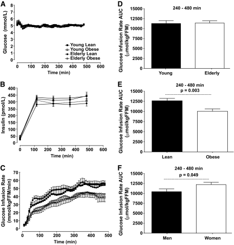
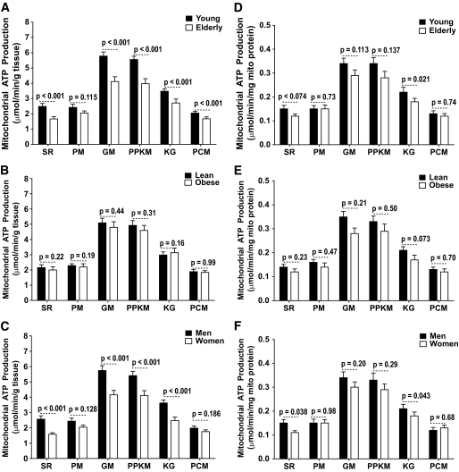
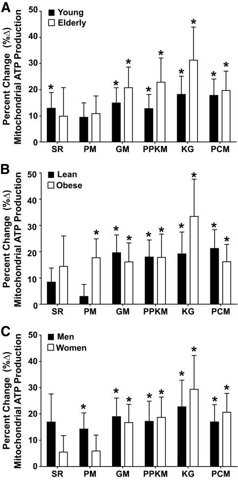
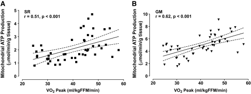
Research design and methods: Twelve young lean, 12 young obese, 12 elderly lean, and 12 elderly obese sedentary adults were studied. Insulin sensitivity was measured by a hyperinsulinemic-euglycemic clamp, and skeletal muscle mitochondrial ATP production rates (MAPRs) were measured in freshly isolated mitochondria obtained from vastus lateralis biopsy samples using the luciferase reaction.

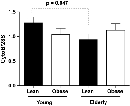
Results: Obese participants, independent of age, had reduced insulin sensitivity based on lower rates of glucose infusion during a hyperinsulinemic-euglycemic clamp. In contrast, age had no independent effect on insulin sensitivity. However, the elderly participants had lower muscle MAPRs than the young participants, independent of obesity. Elderly participants also had higher levels inflammatory cytokines and total adiponectin. In addition, higher muscle MAPRs were also noted in men than in women, whereas glucose infusion rates were higher in women.

Conclusions: The results demonstrate that age-related reductions in insulin sensitivity are likely due to an age-related increase in adiposity rather than a consequence of advanced chronological age. The results also indicate that an age-related decrease in muscle mitochondrial function is neither related to adiposity nor insulin sensitivity. Of interest, a higher mitochondrial ATP production capacity was noted in the men, whereas the women were more insulin sensitive, demonstrating further dissociation between insulin sensitivity and muscle mitochondrial function.


Figures

FIG. 1.
FIG. 1.
Hyperinsulinemic-euglycemic clamp. A and B show the plasma insulin and glucose concentrations during the 8-h clamp, respectively. C shows that the GIRs required to maintain euglycemia during the 8-h clamp were higher in lean groups than in the obese groups with no effects of age. D–F show integrated area under the curve (AUC) for the GIR comparing the young and elderly participants, the lean and obese participants, and men and women, respectively.
FIG. 2.
FIG. 2.
Mitochondrial ATP production rates. Age effect: An age-related decline in mitochondrial ATP production was observed when expressed per gram of tissue using substrates succinate plus rotenone (SR), glutamate plus malate (GM), pyruvate plus palmitoyl-l-carnitine plus α-ketoglutarate plus malate (PPKM), α-ketoglutarate plus glutamate (KG), and palmitoyl-l-carnitine plus 1 malate (PCM) but not pyruvate plus malate (PM) (A). Similar trends were also observed when expressed per milligram of mitochondrial (mito) protein (D). BMI effect: No significant BMI effect was observed when the mitochondrial ATP production rate was expressed per gram of tissue (B) or when expressed per milligram of mitochondrial protein (E). Sex effect: Men had higher MAPRs than women using substrates SR, PM, GM, PPKM, and KG but not PCM when expressed per gram of tissue (C), but significant differences were only observed when using the SR and KG substrates when expressed per milligram of mitochondrial protein content (F). Data are presented as means ± SE.
FIG. 3.
FIG. 3.
Insulin-induced changes in MAPRs. A–C show the percent change (%Δ) in mitochondrial ATP production rates after an 8-h infusion of insulin stratified by age, BMI, and sex, respectively. The 8-h insulin infusion resulted in significant increases in mitochondrial ATP production rates independent of age (A), BMI (B), and sex (C). Data are presented as means ± SE. *P < 0.05 (within group).
FIG. 4.
FIG. 4.
Mitochondrial DNA copy number (mtDNA). Baseline mitochondrial mtDNA copy numbers assessed using primers and probes directed to cytochrome B normalized to 28 s. Data are presented as means ± SE.
FIG. 5.
FIG. 5.
Association between peak Vo2 and mitochondrial ATP production rates. A and B display the relationship between peak Vo2 and mitochondrial ATP production rates assessed using the succinate plus rotenone (SR) and glutamate plus malate (GM) substrate combinations, respectively. Data are presented as means and 95% CIs.

References

    1. Ford ES, Giles WH, Dietz WH: Prevalence of the metabolic syndrome among US adults: findings from the Third National Health and Nutrition Examination Survey. JAMA 2002;287:356–359 - PubMed
    1. Facchini FS, Hua N, Abbasi F, Reaven GM: Insulin resistance as a predictor of age-related diseases. J Clin Endocrinol Metab 2001;86:3574–3578 - PubMed
    1. Short KR, Bigelow ML, Kahl J, Singh R, Coenen-Schimke J, Raghavakaimal S, Nair KS: Decline in skeletal muscle mitochondrial function with aging in humans. Proc Natl Acad Sci U S A 2005;102:5618–5623 - PMC - PubMed
    1. Short KR, Vittone JL, Bigelow ML, Proctor DN, Rizza RA, Coenen-Schimke JM, Nair KS: Impact of aerobic exercise training on age-related changes in insulin sensitivity and muscle oxidative capacity. Diabetes 2003;52:1888–1896 - PubMed
    1. Petersen KF, Dufour S, Befroy D, Garcia R, Shulman GI: Impaired mitochondrial activity in the insulin-resistant offspring of patients with type 2 diabetes. N Engl J Med 2004;350:664–671 - PMC - PubMed

Publication types