. 2009 Oct 30;326(5953):688-694.
doi: 10.1126/science.1179700. Epub 2009 Oct 15.

The crystal structure of the ribosome bound to EF-Tu and aminoacyl-tRNA

Affiliations

The crystal structure of the ribosome bound to EF-Tu and aminoacyl-tRNA

T Martin Schmeing et al. Science. .

Abstract

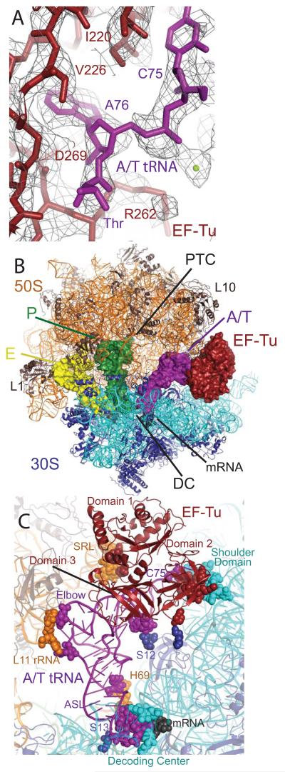
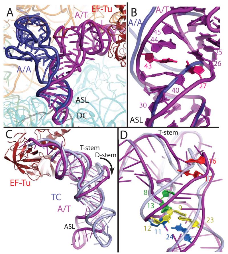
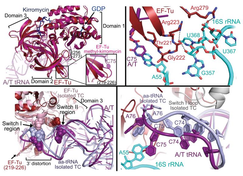
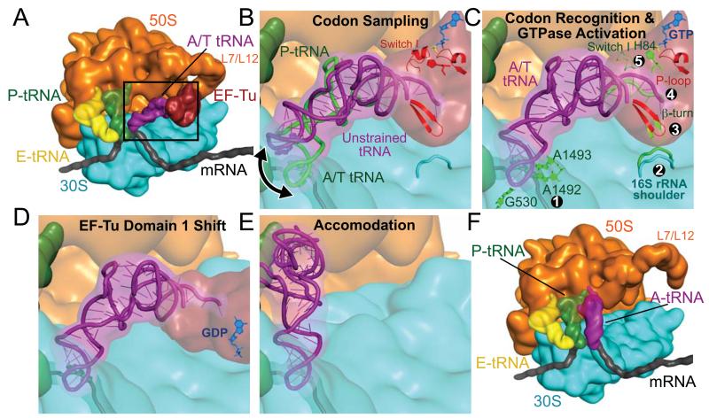
The ribosome selects a correct transfer RNA (tRNA) for each amino acid added to the polypeptide chain, as directed by messenger RNA. Aminoacyl-tRNA is delivered to the ribosome by elongation factor Tu (EF-Tu), which hydrolyzes guanosine triphosphate (GTP) and releases tRNA in response to codon recognition. The signaling pathway that leads to GTP hydrolysis upon codon recognition is critical to accurate decoding. Here we present the crystal structure of the ribosome complexed with EF-Tu and aminoacyl-tRNA, refined to 3.6 angstrom resolution. The structure reveals details of the tRNA distortion that allows aminoacyl-tRNA to interact simultaneously with the decoding center of the 30S subunit and EF-Tu at the factor binding site. A series of conformational changes in EF-Tu and aminoacyl-tRNA suggests a communication pathway between the decoding center and the guanosine triphosphatase center of EF-Tu.


Figures

Fig. 1
Fig. 1
Structure of EF-Tu and aminoacyl-tRNA bound to the ribosome. (A) Representative electron density from an unbiased Fo - Fc map, displayed at 1.3σ, with the refined model of EF-Tu (red) and Thr-tRNAThr (purple). (B) Overall view of the complex, with EF-Tu and tRNAs depicted as surfaces, and rRNA and protein as cartoons. (C) Contacts between TC and the ribosome, with interacting residues shown as spheres.
Fig. 2
Fig. 2
Distortion of aminoacyl-tRNA in the A/T state. (A) Comparison of the A/T tRNA (purple) with the fully accommodated canonical A/A tRNA (dark blue) (30) shows the overall extent of the tRNA disortion. (B) The structures diverge in the anticodon stem loop (ASL) with a reduction of helical twist after basepair 30:40, and separation of the phosphate-sugar backbones at nucleotides 25-45 and 26-44. Disruption of the 27:43 base pair (pink) (9) would facilitate this strand separation. (C) Comparison of the A/T tRNA with tRNA of the isolated TC (light blue) (22) highlights the swinging out and ~5Å shift of D-arm nucleotides. (D) The distortion rationalizes data pertaining to proflavin insertions at nucleotides 16-17 (bright red) (1), cross-linking of nucleotides 8 and 13 (green) (31), and mutations of the Hirsh base pair 24:11 (aqua) (6, 8) and 9:12:23 triple (yellow) (8).
Fig. 3
Fig. 3
Changes of EF-Tu upon ribosome binding. (A) EF-Tu bound to the ribosome (red) adopts a similar conformation to the isolated EF-Tu methyl-kirromycin complex (hot pink)(21), but ribosome binding induces a shift in domain 2 loops including residues 256-273 and 219-226 (inset). (B) The detailed interactions between EF-Tu domain 2 and the shoulder domain of 16S rRNA (cyan). (C) Ribosome binding alters some of the contacts between EF-Tu and tRNA. Interaction of domain 3 with the T-stem is largely unchanged when compared to the isolated TC (22) but that of switch II with the 5′ end of tRNA is altered, and the switch I contact with the 3′ end is abolished. (D) Interaction with the 30S shoulder distorts the 3′ end of tRNA, separating it from switch I, which becomes disordered. The ordered switch I from the isolated TC (grey) is shown as comparison.
Fig. 4
Fig. 4
Schematic representation of the decoding pathway. (A) The L7/L12 stalk recruits TC to a ribosome with deacylated tRNA in the E site and peptidyl-tRNA in the P site. The black frame represents the enlarged area in panels (B)-(E). (B) The tRNA samples codon:anticodon pairing until a match (C) is sensed, by decoding center nucleotides 530 and 1492-3 ①. Codon recognition triggers domain closure of the 30S subunit ②, bringing the shoulder domain into contact with EF-Tu, and shifting the β-loop at 230-237 of domain 2 ③. This changes the conformation of the acceptor end of tRNA ④, disrupting its contacts with switch I, which becomes disordered ⑤, opening the hydrophobic gate to allow His84 to catalyze GTP hydrolysis. (D) GTP hydrolysis and Pi release cause domain rearrangement of EF-Tu, leading to its release from the ribosome and (E-F) accommodation of aminoacyl-tRNA.

Comment in

References

    1. Rodnina MV, Pape T, Fricke R, Kuhn L, Wintermeyer W. J Biol Chem. 1996;271:646. - PubMed
    1. Ogle JM, et al. Science. 2001;292:897. - PubMed
    1. Gromadski KB, Rodnina MV. Mol Cell. 2004;13:191. - PubMed
    1. Ogle JM, Murphy FV, Tarry MJ, Ramakrishnan V. Cell. 2002;111:721. - PubMed
    1. Xia T, et al. Biochemistry. 1998;37:14719. - PubMed

Publication types