. 2009 Oct 23;326(5952):592-6.
doi: 10.1126/science.1178310. Epub 2009 Oct 15.

PTPsigma is a receptor for chondroitin sulfate proteoglycan, an inhibitor of neural regeneration

Affiliations

PTPsigma is a receptor for chondroitin sulfate proteoglycan, an inhibitor of neural regeneration

Yingjie Shen et al. Science. .

Abstract

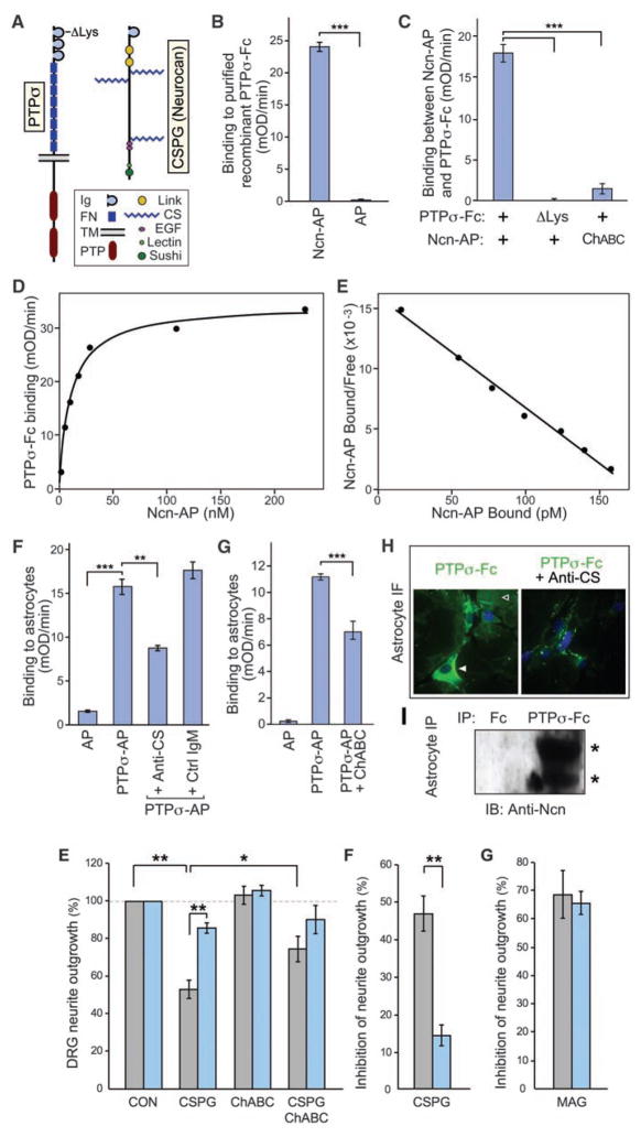
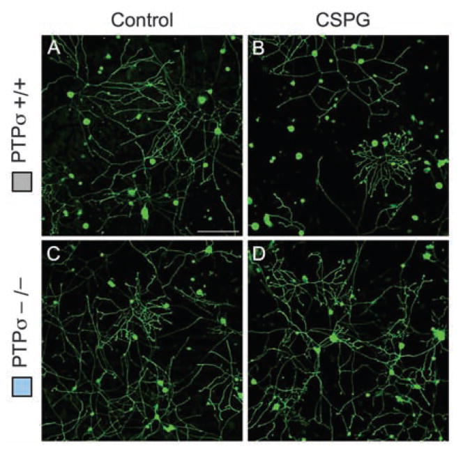
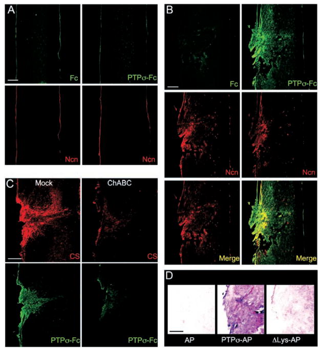
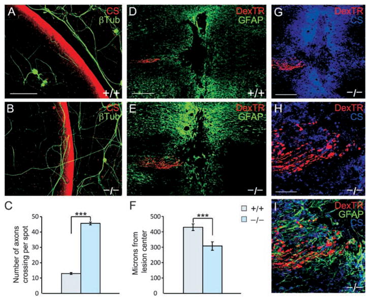
Chondroitin sulfate proteoglycans (CSPGs) present a barrier to axon regeneration. However, no specific receptor for the inhibitory effect of CSPGs has been identified. We showed that a transmembrane protein tyrosine phosphatase, PTPsigma, binds with high affinity to neural CSPGs. Binding involves the chondroitin sulfate chains and a specific site on the first immunoglobulin-like domain of PTPsigma. In culture, PTPsigma(-/-) neurons show reduced inhibition by CSPG. A PTPsigma fusion protein probe can detect cognate ligands that are up-regulated specifically at neural lesion sites. After spinal cord injury, PTPsigma gene disruption enhanced the ability of axons to penetrate regions containing CSPG. These results indicate that PTPsigma can act as a receptor for CSPGs and may provide new therapeutic approaches to neural regeneration.


Figures

Fig. 1
Fig. 1
Binding of the PTPσ ectodomain to CSPG. (A) Domain structure of PTPσ and the CSPG neurocan. ΔLys indicates the site in PTPσ where a cluster of lysines was mutated. (B to E) Purified recombinant PTPσ-Fc fusion protein was immobilized and treated with Ncn-AP fusion protein or AP tag control, followed by quantitation of bound AP activity. (B) Ncn-AP bound above control levels. (C) The PTPσ ΔLys mutation reduced binding to background levels. Pretreatment of Ncn-AP with chondroitinase ABC (ChABC) reduced binding. (D) Binding between PTPσ-Fc and Ncn-AP was saturable. (E) Scatchard analysis produced a linear plot, indicating a single binding affinity with KD = 11 nM. (F) PTPσ-AP bound to C8-D1A astrocyte cultures above AP control levels. Binding was reduced by antibody to CS (anti-CS), but not by an immunoglobulin M (IgM) control. (G) Chondroitinase ABC pretreatment of astrocyte cultures reduced PTPσ-AP binding. (H) PTPσ-Fc immunofluorescence (green) showed binding over cell surfaces (solid arrowhead) and extracellular matrix (open arrowhead); Fc control gave no visible fluorescence (not shown in the figure). Pre-incubation with anti-CS reduced binding. Blue is nuclear counter-stain. (I) PTPσ-Fc, but not Fc control, coprecipitated neurocan from C8-D1A astrocyte lysates. Western blot showed bands with expected sizes for neurocan proteolytic fragments at approximately 150 and 100 kD (asterisks). ***P < 0.001, **P < 0.01.
Fig. 2
Fig. 2
Effect of PTPσ deficiency on the response of sensory neurons to CSPG. (A to D) DRG neurons from P8 mice were grown for 18 hours, then treated for 24 hours with or without CSPG and visualized by GAP-43 immunolabeling. (E) Quantitation of neurite outgrowth. PTPσ−/− neurons showed significantly less inhibition by CSPG than wild-type neurons did. A significant difference was no longer seen when chondroitinase ABC was added along with the CSPG. (F and G) Effect of CSPG or MAG, expressed as percentage of inhibition of outgrowth. PTPσ deficiency reduced the inhibitory action of CSPG but had no significant effect on the inhibitory action of MAG. n = 5 mice for each genotype. *P < 0.05, **P < 0.01. Scale bars, 100 μm.
Fig. 3
Fig. 3
PTPσ fusion protein detection of ligand distribution at a spinal cord lesion site. (A and B) Sections were double–fluorescence-labeled with antibody to neurocan (Ncn, red), together with either PTPσ-Fc probe (green) or Fc control (adjacent section, green). (A) Unlesioned spinal cord. Anti-neurocan labeled a thin line at the pia. PTPσ-Fc showed no labeling noticeably above Fc control levels. (B) Spinal cord 7 days after dorsal hemisection. The lesion site showed simultaneous elevation of neurocan immunolabeling and PTPσ-Fc binding. The distributions overlapped, with PTPσ-Fc labeling additional areas. (C) ChABC treatment reduced binding of PTPσ-Fc to the lesion site. Adjacent sections were preincubated with or without ChABC, then double-labeled with anti-CS (red) and PTPσ-Fc (green). (D) Introduction of ΔLys mutation into a PTPσ-AP probe reduced lesion site binding (purple) close to AP tag control levels. Scale bars, 200 μm.
Fig. 4
Fig. 4
Adult PTPσ−/− sensory neurons show reduced sensitivity to inhibition by a proteoglycan gradient and can extend further in a spinal cord lesion. (A and B) Adult wild-type DRG neurons visualized by antibody to β-tubulin (green) avoided the most inhibitory rim of the gradient visualized by anti-CS (red). PTPσ−/− neurons showed greater ability to cross the rim. (C) Quantitation of the average number of axons per spot growing up the gradient and crossing the outer rim. Wild-type, n = 48 spots; PTPσ−/−, n = 40 spots; ***P < 0.001. (D and E) Confocal images of longitudinal sections from adult mouse spinal cord 14 days after dorsal column crush, caudal to the left. Sensory axons are labeled with DexTR (red), and the lesion is delineated by glial fibrillary acidic protein+ (GFAP+) astrocytes (green). Wild-type axons are seen several hundred micrometers from the lesion center; PTPσ−/−axons abut the edge of the lesion core. (F) Quantitation of distance from lesion center. Wild-type, n = 35 mice; PTPσ−/−, n = 30 mice. ***P < 0.002. (G to I) Confocal z-stack images of PTPσ−/− mouse dorsal column crush lesion, showing relationship between injured fibers (red), anti-CS labeling (blue), and in (I), reactive astrocytes (green). Scale bars in (A) and (B), 100 μm; in (D) and (E), 200 μm; in (G) to (I), 50 μm.

References

    1. Case LC, Tessier-Lavigne M. Curr Biol. 2005;15:R749. - PubMed
    1. Domeniconi M, Filbin MT. J Neurol Sci. 2005;233:43. - PubMed
    1. Liu BP, Cafferty WB, Budel SO, Strittmatter SM. Philos Trans R Soc London Ser B. 2006;361:1593. - PMC - PubMed
    1. Lu P, Tuszynski MH. Exp Neurol. 2008;209:313. - PMC - PubMed
    1. Yiu G, He Z. Nat Rev Neurosci. 2006;7:617. - PMC - PubMed

MeSH terms