. 2009 Oct 16;326(5951):426-9.
doi: 10.1126/science.1177350.

Generation of functional ventricular heart muscle from mouse ventricular progenitor cells

Affiliations

Generation of functional ventricular heart muscle from mouse ventricular progenitor cells

Ibrahim J Domian et al. Science. .

Abstract

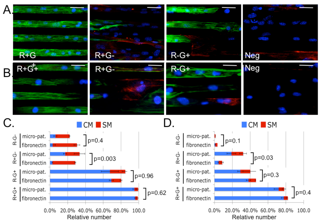
The mammalian heart is formed from distinct sets of first and second heart field (FHF and SHF, respectively) progenitors. Although multipotent progenitors have previously been shown to give rise to cardiomyocytes, smooth muscle, and endothelial cells, the mechanism governing the generation of large numbers of differentiated progeny remains poorly understood. We have employed a two-colored fluorescent reporter system to isolate FHF and SHF progenitors from developing mouse embryos and embryonic stem cells. Genome-wide profiling of coding and noncoding transcripts revealed distinct molecular signatures of these progenitor populations. We further identify a committed ventricular progenitor cell in the Islet 1 lineage that is capable of limited in vitro expansion, differentiation, and assembly into functional ventricular muscle tissue, representing a combination of tissue engineering and stem cell biology.


Figures

Figure 1
Figure 1. ED9.5 SHF-dsRed/Nkx2.5-eGFP transgenic embryos
A-C. Whole mount fluorescence microscopy of double transgenic ED9.5 embryos. The LV and IFT are eGFP+, the RV and OT are dsRed+ and eGFP+, and the PM is dsRed+. Right lateral (A) and (B), and left lateral (C) views are shown. Scale bar 500µm (A) and 100µm (B and C). D. Immunofluorescence labeling of ED9.5 double transgenic mouse embryo shows that cells in the RV and OFT are R+G+, in the PM are R+G-, and in the LV and IFT are R-G+. Section levels are shown in cartoon. eGFP is shown in green, dsRed in Red, and overlay in yellow. Scale bar 50µm.
Figure 2
Figure 2. Isolation of cardiac progenitors
A&B. Representative flow cytometry plots of double transgenic day 6 EBs (A) and ED9.5 embryos (B) showing 4 populations of cells: R+G+, R+G-, RG+, and R-G-. C. qPCR analysis of mRNA and miRNA isolated from embryonic progenitors. Values are normalized against unlabeled control. Standard deviation (SD) shown. Differences between groups were highly statistically significant, see Table S2 and S3. D. Hoechst staining and FACS-based cell cycle analysis of undifferentiated ESC, EB day6 progenitors, and differentiated progeny. E. ESC derived cardiac progenitors were cultured 5 days. DAPI and Ki67 staining was performed to quantify total cell number and proportion of cycling cells (Ki67+ cells/total cells). SD shown (n=4).
Figure 3
Figure 3. Differentiation potential of cardiac progenitors
Embryonic and ESC-derived cardiac progenitors were cultured on fibronectin coated slides (fibronectin) or micro-patterns for five days. A&B. Representative immunofluorescence microscopy of Embryonic (A) or ESC-derived (B) progenitors cultured on micropatterns are shown; nuclei (blue), smMHC (red) and sarcomeric α-actinin (green). Scale bar 40µm. C&D. Cell counting was used to quantify the relative number of CM (sarcomeric α-actinin positive) or SM (smMHC positive) derived from embryonic (C) or ESC (D) progenitors. R+G+ populations resulted in the most CM (p<0.001). No significant differences were observed in SM differentiation (p=0.38-1.0). P-values for the differences in CM differentiation are displayed.
Figure 4
Figure 4. Engineered ventricular tissue from R+G+ progenitors
A. R+G+ (n=12), R+G- (n=5), and R-G+ (n=5) progenitors were allowed to differentiate and single cell patch clamp recordings were performed. AP morphology was assessed for typical four-phase ventricular action potential. B. Representative spontaneous AP from R+G+ derived cardiomyocytes. C. ESC-derived R+G+ progenitors were used to generate MTF as described in Supplementary Materials and (14). Field stimulation (10 V, 10 ms pulse-width) induced 0.5 Hz cyclical contraction and rhythmic MTF bending. See Movie S1.

References

    1. Buckingham M, Meilhac S, Zaffran S. Nat Rev Genet. 2005 Nov;6:826. - PubMed
    1. Martin-Puig S, Wang Z, Chien KR. Cell Stem Cell. 2008;2:320. - PubMed
    1. Dodou E, Xu SM, Black BL. Mech Dev. 2003 Sep;120:1021. - PubMed
    1. Qyang Y, et al. Cell Stem Cell. 2007;1:165. - PubMed
    1. Lien CL, et al. Development. 1999 Jan;126:75. - PubMed

Publication types

MeSH terms

Associated data