

Biology of vascular malformations of the brain
- PMID: 19834013
- PMCID: PMC2810509
- DOI: 10.1161/STROKEAHA.109.563692
Biology of vascular malformations of the brain
Abstract
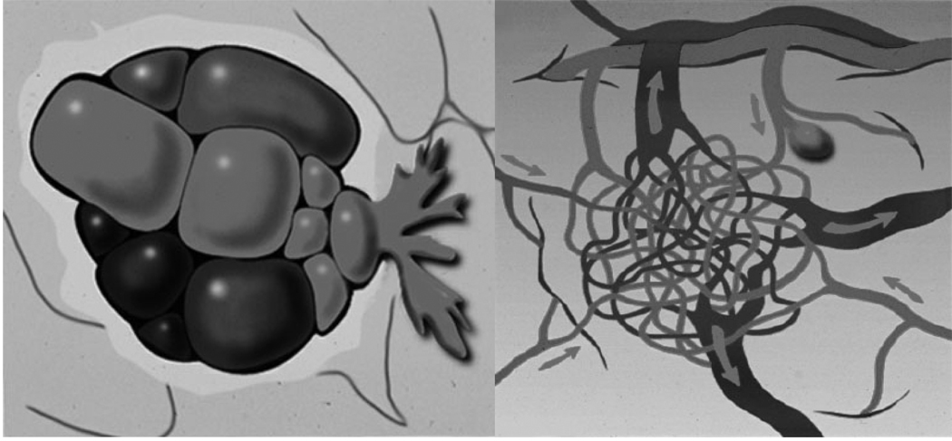
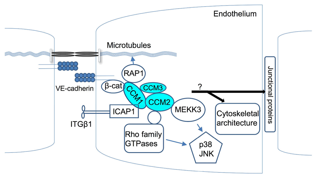
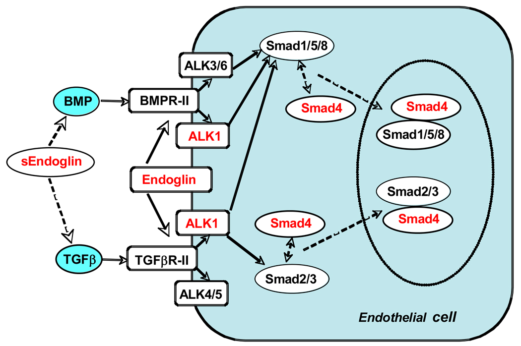
Background and purpose: This review discusses recent research on the genetic, molecular, cellular, and developmental mechanisms underlying the etiology of vascular malformations of the brain (VMBs), including cerebral cavernous malformation, sporadic brain arteriovenous malformation, and the arteriovenous malformations of hereditary hemorrhagic telangiectasia. Summary of Review- The identification of gene mutations and genetic risk factors associated with cerebral cavernous malformation, hereditary hemorrhagic telangiectasia, and sporadic arteriovenous malformation has enabled the development of animal models for these diseases and provided new insights into their etiology. All of the genes associated with VMBs to date have known or plausible roles in angiogenesis and vascular remodeling. Recent work suggests that the angiogenic process most severely disrupted by VMB gene mutation is that of vascular stabilization, the process whereby vascular endothelial cells form capillary tubes, strengthen their intercellular junctions, and recruit smooth muscle cells to the vessel wall. In addition, there is now good evidence that in some cases, cerebral cavernous malformation lesion formation involves a genetic 2-hit mechanism in which a germline mutation in one copy of a cerebral cavernous malformation gene is followed by a somatic mutation in the other copy. There is also increasing evidence that environmental second hits can produce lesions when there is a mutation to a single allele of a VMB gene.
Conclusions: Recent findings begin to explain how mutations in VMB genes render vessels vulnerable to rupture when challenged with other inauspicious genetic or environmental factors and have suggested candidate therapeutics. Understanding of the cellular mechanisms of VMB formation and progression in humans has lagged behind that in animal models. New knowledge of lesion biology will spur new translational work. Several well-established clinical and genetic database efforts are already in place, and further progress will be facilitated by collaborative expansion and standardization of these.
Figures
Similar articles
-
Cerebral cavernous malformations as a disease of vascular permeability: from bench to bedside with caution.Neurosurg Focus. 2010 Sep;29(3):E4. doi: 10.3171/2010.5.FOCUS10121. Neurosurg Focus. 2010. PMID: 20809762 Free PMC article. Review.
-
Arteriovenous malformations and other vascular malformation syndromes.Cold Spring Harb Perspect Med. 2013 Feb 1;3(2):a006635. doi: 10.1101/cshperspect.a006635. Cold Spring Harb Perspect Med. 2013. PMID: 23125071 Free PMC article. Review.
-
Systemic and CNS manifestations of inherited cerebrovascular malformations.Clin Imaging. 2021 Jul;75:55-66. doi: 10.1016/j.clinimag.2021.01.020. Epub 2021 Jan 20. Clin Imaging. 2021. PMID: 33493737 Free PMC article. Review.
-
Deregulated TGF-β/BMP Signaling in Vascular Malformations.Circ Res. 2017 Sep 29;121(8):981-999. doi: 10.1161/CIRCRESAHA.117.309930. Circ Res. 2017. PMID: 28963191 Review.
-
Recent Advances in Basic Research for Brain Arteriovenous Malformation.Int J Mol Sci. 2019 Oct 25;20(21):5324. doi: 10.3390/ijms20215324. Int J Mol Sci. 2019. PMID: 31731545 Free PMC article. Review.
Cited by
-
NIR-II window tracking of hyperglycemia induced intracerebral hemorrhage in cerebral cavernous malformation deficient mice.Biomater Sci. 2020 Sep 21;8(18):5133-5144. doi: 10.1039/d0bm00873g. Epub 2020 Aug 21. Biomater Sci. 2020. PMID: 32821891 Free PMC article.
-
Excess vascular endothelial growth factor-A disrupts pericyte recruitment during blood vessel formation.Angiogenesis. 2019 Feb;22(1):167-183. doi: 10.1007/s10456-018-9648-z. Epub 2018 Sep 20. Angiogenesis. 2019. PMID: 30238211 Free PMC article.
-
LINE-1 retrotransposition events affect endothelial proliferation and migration.Histochem Cell Biol. 2010 Dec;134(6):581-9. doi: 10.1007/s00418-010-0758-y. Epub 2010 Nov 11. Histochem Cell Biol. 2010. PMID: 21069374
-
Cerebral arteriovenous malformation presenting with polycythemia vera: A case report and literature review.Int J Surg Case Rep. 2013;4(10):813-7. doi: 10.1016/j.ijscr.2013.06.019. Epub 2013 Jul 20. Int J Surg Case Rep. 2013. PMID: 23959406 Free PMC article.
-
PTEN/PI3K/Akt/VEGF signaling and the cross talk to KRIT1, CCM2, and PDCD10 proteins in cerebral cavernous malformations.Neurosurg Rev. 2015 Apr;38(2):229-36; discussion 236-7. doi: 10.1007/s10143-014-0597-8. Epub 2014 Nov 19. Neurosurg Rev. 2015. PMID: 25403688 Review.
References
-
- Al-Shahi R, Bhattacharya JJ, Currie DG, Papanastassiou V, Ritchie V, Roberts RC, Sellar RJ, Warlow CP. Prospective, population-based detection of intracranial vascular malformations in adults: The scottish intracranial vascular malformation study (sivms) Stroke. 2003;34:1163–1169. - PubMed
-
- Plate KH. Mechanisms of angiogenesis in the brain. J Neuropathol Exp Neurol. 1999;58:313–320. - PubMed
-
- Greenberg DA, Jin K. From angiogenesis to neuropathology. Nature. 2005;438:954–959. - PubMed
-
- Vorbrodt AW, Dobrogowska DH, Tarnawski M. Immunogold study of interendothelial junction-associated and glucose transporter proteins during postnatal maturation of the mouse blood-brain barrier. J Neurocytol. 2001;30:705–716. - PubMed
Publication types
MeSH terms
Grants and funding
LinkOut - more resources
Full Text Sources
Other Literature Sources
