. 2009 Nov;175(5):2053-62.
doi: 10.2353/ajpath.2009.090211. Epub 2009 Oct 15.

Glycosaminoglycan-mediated loss of cathepsin K collagenolytic activity in MPS I contributes to osteoclast and growth plate abnormalities

Affiliations

Glycosaminoglycan-mediated loss of cathepsin K collagenolytic activity in MPS I contributes to osteoclast and growth plate abnormalities

Susan Wilson et al. Am J Pathol. 2009 Nov.

Abstract

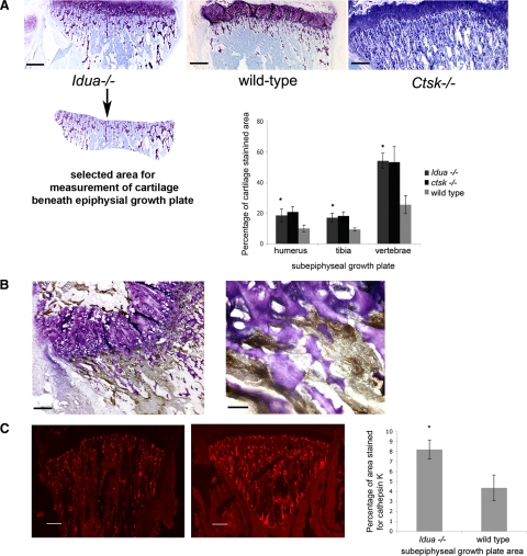
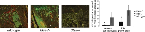
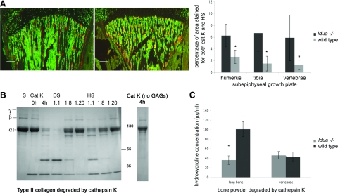
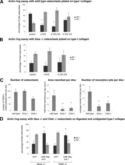
Mucopolysaccharidoses are a group of lysosomal storage diseases characterized by the build-up of glycosaminoglycans (GAGs) and severe skeletal abnormalities. As GAGs can regulate the collagenolytic activity of the major osteoclastic protease cathepsin K, we investigated the presence and activity of cathepsin K and its co-localization with GAGs in mucopolysaccharidosis (MPS) type I bone. The most dramatic difference between MPS I and wild-type mice was an increase in the amount of cartilage in the growth plates in MPS I bones. Though the number of cathepsin K-expressing osteoclasts was increased in MPS I mice, these mice revealed a significant reduction in cathepsin K-mediated cartilage degradation. As excess heparan and dermatan sulfates inhibit type II collagen degradation by cathepsin K and the spatial overlap between cathepsin K and heparan sulfate strongly increased in MPS I mice, the build up of subepiphyseal cartilage is speculated to be a direct consequence of cathepsin K inhibition by MPS I-associated GAGs. Moreover, isolated MPS I and Ctsk(-/-) osteoclasts displayed fewer actin rings and formed fewer resorption pits on dentine disks, as compared with wild-type cells. These results suggest that the accumulation of GAGs in murine MPS I bone has an inhibitory effect on cathepsin K activity, resulting in impaired osteoclast activity and decreased cartilage resorption, which may contribute to the bone pathology seen in MPS diseases.


Figures

Figure 1
Figure 1
A: Toluidine blue staining of the tibial growth plate area in Idua−/−, wild-type (WT), and Ctsk−/− mice. Graph showing percentage of cartilage area in the subepiphyseal growth plate of shoulder and knee joints, as well as vertebra bones. *P < 0.05 vs wild-type. The panel below the Idua−/− growth plate represents an example for the measured cartilage-containing area (in all sections a constant area was analyzed: about 700,00 μm2 in tibia). Scale bars = 260 μm. B: Diaminobenzidine staining of cathepsin K as an osteoclast marker in subepyphysial growth plate area. The asterisk in the left panel represents the area in the right panel at a higher magnification (magnification bars are 130 μm and 40 μm, respectively). Cathepsin K-expressing cells are clearly seen adjacent to toluidine-blue stained cartilage. C: Cathepsin K staining of the subepiphyseal growth plate area in tibia of wild-type and Idua−/− mice. Graph showing the average percentage of subepiphyseal area stained positive for cathepsin K in shoulder, knee joints, and vertebra bones for wild-type and Idua−/−. *P < 0.05 vs. wild-type. Scale bars = 260 μm.
Figure 2
Figure 2
Neoepitope staining of cathepsin K catalyzed type II collagen cleavage in subepiphyseal growth plate area of wild-type (WT) and Idua−/− tibia (red or yellow for neoepitopes; green is enhanced background using the green filter to visualize the joint structure). Asterisks indicate sporadic artifact fluorescent precipitations). Graph showing percentage of area beneath growth plate containing cleaved type II collagen staining in humerus (shoulder) and tibia (knee) joints of wild-type, idua−/−, and ctsk−/− mice. *P < 0.05 vs. wild-type.
Figure 3
Figure 3
A: Co-localization (orange) of heparan sulfate (green) and cathepsin K (red) in the growth plate of tibia in wild-type (WT) and Idua−/− mice. Graph of percentage of overlap in total subgrowth plate area of humerus, tibia, and vertebrae. *P < 0.05 vs wild-type. Scale bars = 260 μm. B: SDS-polyacrylamide gel electrophoresis of type II collagen degradation at a 1:1 ratio of cathepsin K (800 nmol/L) and chondroitin sulfate (800 nmol/L) (CS) and in the presence of additional and increasing amounts of MPS1-related GAGs (DS or HS) added at final ratios of cathepsin K to DS or HS at 1:1, 1:8, and 1:20. Reactions were in sodium acetate buffer for 4 hours at 28°C. C: Hydroxyproline assay: long bone and vertebrae bone powder from Idua−/− and wild-type bone were crushed and subject to digestion by 800 nmol/L cathepsin K in sodium acetate buffer overnight at 28°C. The soluble fragments were assayed for hydroxyproline against known standards. *P < 0.05 vs. wild-type.
Figure 4
Figure 4
Osteoclast cultures obtained from murine long bones were seeded on type I collagen substrate, after 24 hours incubation cultures were stained for actin with FITC-phalloidin. Osteoclasts displaying a full actin ring or disrupted actin rings with more than 50% intact were identified as active. Osteoclasts displaying a disrupted actin ring (diffuse ring or less than 50% intact ring), or complete lack of actin structure, were identified as inactive. After 24 hours incubation, cultures were stained for actin and the total number of osteoclasts with actin rings were counted and compared with the total number of osteoclasts present (determined by cells containing two or more nuclei). Bar chart shows comparison between percentage of osteoclasts displaying an actin ring (positive for Actin Ring +) or without (negative for Actin Ring −) under different experimental conditions. Data are expressed as percentage. A: Osteoclasts were incubated with or without 5 μmol/L of the broad spectrum cathepsin inhibitor, LHVS. Osteoclasts were also incubated in the presence of 0.15% dermatan sulfate (DS) or chondroitin sulfate (CS), GAGs were incorporated in the type I collagen matrix and the cell media. After 24 hours incubation, cultures were stained for actin and the total number of osteoclasts with actin rings were counted and compared with the total number of osteoclasts. *P < 0.05 vs wild-type untreated control. B: Idua−/− osteoclasts were incubated in the presence and absence of 5 μmol/L of the broad spectrum cathepsin inhibitor, LHVS, and 0.15% dermatan sulfate in the matrix and media. C: Wild-type, Idua−/− and Ctsk−/− osteoclasts were cultured on dentine disks for 48 hours. The number of TRAP+ osteoclasts and the number and area of resorption pits were analyzed. Values are the means plus SD of four dentine slices. *P < 0.05 vs. wild-type. D: Long bone osteoclasts obtained from Idua−/− and Ctsk−/− mice were seeded onto intact type I collagen matrix (coll 1) or type I collagen pre-degraded by cathepsin K (catK deg coll 1) and analyzed for actin ring presence. Collagen type I was pre-degraded by 400 nmol/L cathepsin K in the presence of chondroitin 4-sulfate for 4 hours at 28°C. After 24 hours, cultures were stained for actin and the percentage of osteoclasts displaying an actin ring (AR+) is shown compared with the percentage without (AR−). *P ≤ 0.05 when compared with same osteoclasts cultured on intact collagen matrix.

References

    1. Neufeld EF, Muenzer J. The Mucopolysaccharidoses. New York: McGraw-Hill,; 2001:pp 3419–3428.
    1. Burrow TA, Hopkin RJ, Leslie ND, Tinkle BT, Grabowski GA. Enzyme reconstitution/replacement therapy for lysosomal storage diseases. Curr Opin Pediatr. 2007;19:628–635. - PubMed
    1. Yasuda Y, Kaleta J, Bromme D. The role of cathepsins in osteoporosis and arthritis: rationale for the design of new therapeutics. Adv Drug Deliv Rev. 2005;57:973–993. - PubMed
    1. Gelb BD, Shi GP, Chapman HA, Desnick RJ. Pycnodysostosis, a lysosomal disease caused by cathepsin K deficiency. Science. 1996;273:1236–1238. - PubMed
    1. Everts V, Hou WS, Rialland X, Tigchelaar W, Saftig P, Bromme D, Gelb BD, Beertsen W. Cathepsin K deficiency in pycnodysostosis results in accumulation of non-digested phagocytosed collagen in fibroblasts. Calcif Tissue Int. 2003;73:380–386. - PubMed

Publication types