. 2009 Nov;175(5):2146-58.
doi: 10.2353/ajpath.2009.090218. Epub 2009 Oct 15.

Cripto-1 is required for hypoxia to induce cardiac differentiation of mouse embryonic stem cells

Affiliations

Cripto-1 is required for hypoxia to induce cardiac differentiation of mouse embryonic stem cells

Caterina Bianco et al. Am J Pathol. 2009 Nov.

Abstract

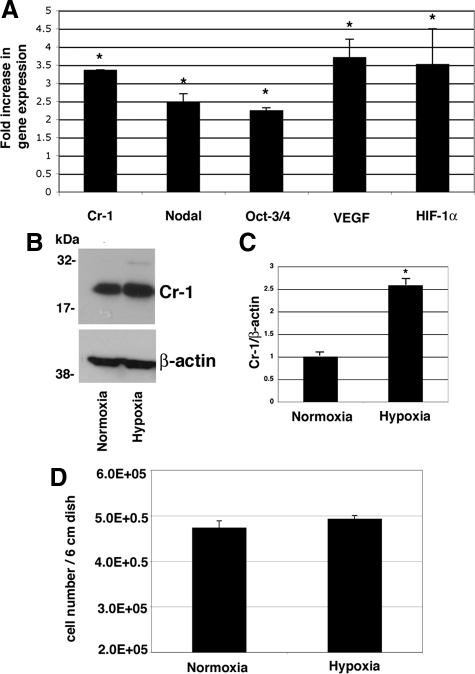
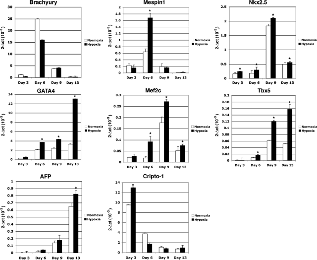
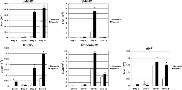
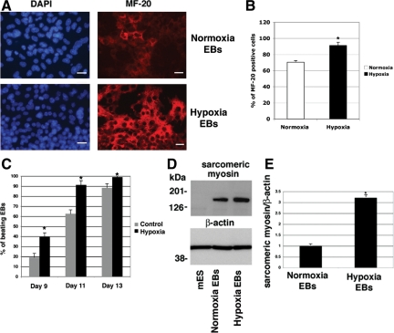
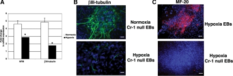
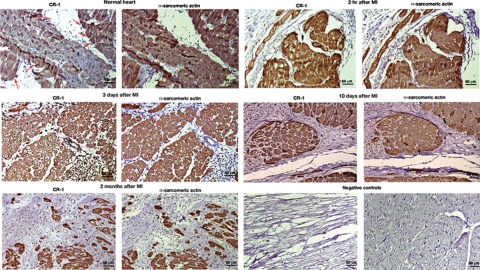
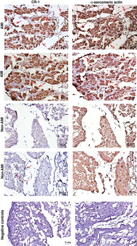
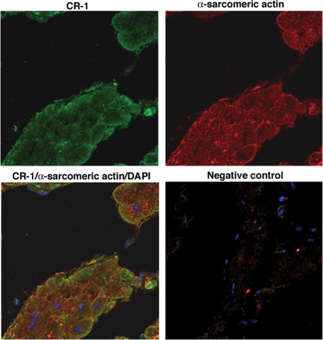
Cripto-1 is a membrane-bound protein that is highly expressed in embryonic stem cells and in human tumors. In the present study, we investigated the effect of low levels of oxygen, which occurs naturally in rapidly growing tissues, on Cripto-1 expression in mouse embryonic stem (mES) cells and in human embryonal carcinoma cells. During hypoxia, Cripto-1 expression levels were significantly elevated in mES cells and in Ntera-2 or NCCIT human embryonal carcinoma cells, as compared with cells growing with normal oxygen levels. The transcription factor hypoxia-inducible factor-1alpha directly regulated Cripto-1 expression by binding to hypoxia-responsive elements within the promoter of mouse and human Cripto-1 genes in mES and NCCIT cells, respectively. Furthermore, hypoxia modulated differentiation of mES cells by enhancing formation of beating cardiomyocytes as compared with mES cells that were differentiated under normoxia. However, hypoxia failed to induce differentiation of mES cells into cardiomyocytes in the absence of Cripto-1 expression, demonstrating that Cripto-1 is required for hypoxia to fully differentiate mES cells into cardiomyocytes. Finally, cardiac tissue samples derived from patients who had suffered ischemic heart disease showed a dramatic increase in Cripto-1 expression as compared with nonischemic heart tissue samples, suggesting that hypoxia may also regulate Cripto-1 in vivo.


Figures

Figure 1
Figure 1
Hypoxia up-regulates Cr-1 mRNA and protein expression in mES cells. A: mES cells were grown in a hypoxic chamber for 24 hours, and RNA was extracted and analyzed by real-time PCR. Data were normalized to 28S expression and presented as fold increase versus the corresponding mRNA level in mES growing with normal oxygen levels. Data are representative of five experiments with triplicate samples. P < 0.05. B: Western blot analysis for Cr-1 in mES cells growing in normoxia or hypoxia for 24 hours. C: Densitometric analysis of Cr-1 in mES cell lysates normalized to β-actin content. Densitometric analysis is representative of three Western blot experiments. P < 0.05. D: Proliferation assay of mES cells growing in normoxia or hypoxia for 24 hours. Data are representative of three experiments with duplicate samples.
Figure 2
Figure 2
The HIF-1α inhibitors, echinomycin and emetine, interfere with induction of Cr-1 protein expression by hypoxia in mES cells. A: mES cells were incubated in a hypoxic chamber in the presence or absence of various concentrations of echinomycin (5 or 2.5 nmol/L) or emetine (0.25 or 0.5 μmol/L) for 24 hours and analyzed by Western blot using anti-Cr-1 or anti-β-actin antibodies. B: Fold difference in Cr-1 protein expression by desitometric analysis of Cr-1 in mES cell lysates normalized to β-actin content. Densitometric analysis is representative of three Western blot experiments. P < 0.05, as compared with hypoxia-stimulated nontreated cells.
Figure 3
Figure 3
Hypoxia up-regulates Cr-1 mRNA and protein expression through HIF-1α. Real-time PCR for HIF-1α (A) and Cr-1 (B) in mES cells transfected with HIF-1α or control siRNAs. mRNA expression was normalized to 28S expression. Data are representative of three experiments with duplicate samples. P < 0.05, as compared with hypoxia-stimulated nontransfected cells. C: Western blot analysis for Cr-1 and β-actin in siRNA transfected mES cells. D: Fold difference in Cr-1 protein expression by desitometric analysis of Cr-1 in mES cell lysates normalized to β-actin content. Densitometric analysis is representative of three Western blot experiments. P < 0.05, as compared with hypoxia-stimulated nontransfected cells.
Figure 4
Figure 4
HIF-1α directly binds to the mouse and human Cripto-1 promoter in mES and NCCIT cells and enhances CR-1 promoter luciferase activity in COS7 cells. A: CHIP assays in mES and NCCIT cells following 24 hours of incubation in a hypoxic chamber. The cross-linked protein-DNA complexes were immunoprecipitated with anti-HIF-1α antibody or with an isotype control IgG as negative control, and the purified DNA was amplified by PCR using specific primer sets that amplified the HRE region (HRE1) within the mouse and human Cripto-1 promoters. The input control and hypoxia samples are DNA samples before the immunoprecipitation that were used as loading control for the PCR. M, DNA 100 bp marker. B: Dual-luciferase assay of transiently transfected COS7 cells with a full-length 2.5-kb human CR-1-luciferase promoter. Cells were subsequently treated with the hypoxic mimetics desferrioxamine (DFX) (65, 130, or 260 μmol/L) or CoCl2 (100 or 200 μmol/L) or incubated in a hypoxic chamber for an additional 24 hours. Data are representative of four experiments with duplicate samples. P < 0.001. RLU, relative luciferase units.
Figure 5
Figure 5
Hypoxia enhances mesoderm and cardiac differentiation of EBs. mES cells were differentiated into cardiomyocytes using the hanging drop technique, and RNA samples were collected at days 3, 6, 9, and 13 of differentiation. Hypoxia stimulation of EBs occurred at day 2 of differentiation for 24 hours. Markers were analyzed by real-time PCR, and data were normalized to 28S expression. Data are representative of four experiments with duplicate samples. P < 0.001.
Figure 6
Figure 6
Hypoxia promotes cardiomyocyte differentiation of ES cells enhancing expression of terminal myocardial differentiation genes. Real-time PCR for α-MHC, β-MHC, MLC2v, troponin T2, and atrial natriuretic factor (ANF) in differentiating normoxia or hypoxia stimulated EBs. Data are representative of four experiments with duplicate samples. P < 0.001.
Figure 7
Figure 7
Hypoxia increases sarcomeric myosin expression in EBs. A: Immunofluorescence staining for sarcomeric-myosin using anti-MF-20 monoclonal antibody (red) of normoxia or hypoxia-stimulated EBs at 13 days of differentiation. Nuclear staining is shown in blue (4′,6′-diamidino-2-phenylindole). Scale bar = 20 μm. Magnification is ×40. B: Percentage of MF-20-positive cells/field. Values are averages of number of positive cells counted in several fields (between 5 and 8). P < 0.05. C: Percentage of beating EBs counted from days 9 to 13 of differentiation. For each time point the number of EBs counted were between 35 and 50. Data are representative of three experiments. D: Western blot analysis for sarcomeric-myosin in normoxia or hypoxia stimulated EBs at day 13 of differentiation using the MF-20 antibody. Membranes were reprobed with an anti-β-actin antibody and densitometrically analyzed (E) by normalization of sarcomeric-myosin levels to β-actin content. Densitometric analysis is representative of two Western blot experiments. P < 0.05.
Figure 8
Figure 8
Hypoxia interferes with neuronal differentiation of Cr-1−/− EBs. A: Real-time PCR for neuronal markers NFM and βIII-tubulin in normoxia or hypoxia-stimulated Cr-1−/− EBs at 13 days of differentiation. Data were normalized to 28S content. Data are representative of two experiments with triplicate samples. P < 0.001. B: Immunofluorescence staining of βIII-tubulin (green) in normoxia or hypoxia stimulated Cr-1−/− EBs at 13 days of differentiation. 4′,6′-Diamidino-2-phenylindole staining of nuclei is shown in blue. Scale bar = 20 μm. Magnification is ×40. C: Immunofluorescence staining for MF-20 in hypoxia-stimulated wild-type and Cr-1−/− EBs. Magnification is ×20.
Figure 9
Figure 9
Hypoxia enhances mesodermal and cardiac gene expression in Cr-1−/− EBs but it is not sufficient to induce cardiomyocyte differentiation. Real-time PCR for mesodermal and cardiac markers in normoxia or hypoxia stimulated Cr-1−/− EBs at different time points during differentiation. Data were normalized to 28S expression. Data are representative of two experiments with triplicate samples. P < 0.001.
Figure 10
Figure 10
Cripto-1 cardiac expression in an animal model of MI. Immunohistochemistry for CR-1 and α-sarcomeric actin in serial heart tissue sections of a porcine model of myocardial ischemia-reperfusion. Heart tissue samples were collected after 2 hours, 3 days, 10 days, and 2 months of induced MI. A normal heart sample was harvested from a healthy pig. Red arrows in the normal heart are pointing to some of the cells that show positive staining for CR-1. Negative controls were obtained by omitting the primary antibodies only. Scale bar = 50 μm. Original magnifications, ×20.
Figure 11
Figure 11
CR-1 expression is increased in human AMI. Immunohistochemistry for CR-1 and α-sarcomeric actin in AMI and non-AMI human heart serial tissue sections. The figure shows two different patients with AMI and two different patients with non-AMI. Red arrows in AMI samples are pointing to some of the cells that are positive for both CR-1 and α-sarcomeric actin. Negative controls were obtained by omitting the primary antibodies only. Scale bar = 50 μm. Original magnification, ×20.
Figure 12
Figure 12
Immunofluorescent analysis for CR-1 and α-sarcomeric actin in heart frozen tissue section from patients with ischemic heart disease. Negative control was obtained by incubating the slides with secondary antibodies only. Fluorescence images were acquired by laser scanning confocal microscopy.

References

    1. Bianco C, Strizzi L, Normanno N, Khan N, Salomon DS. Cripto-1: an oncofetal gene with many faces. Curr Top Dev Biol. 2005;67:85–133. - PubMed
    1. Schier AF. Nodal signaling in vertebrate development. Annu Rev Cell Dev Biol. 2003;19:589–621. - PubMed
    1. Dono R, Scalera L, Pacifico F, Acampora D, Persico MG, Simeone A. The murine cripto gene: expression during mesoderm induction and early heart morphogenesis. Development. 1993;118:1157–1168. - PubMed
    1. Xu C, Liguori G, Persico MG, Adamson ED. Abrogation of the Cripto gene in mouse leads to failure of postgastrulation morphogenesis and lack of differentiation of cardiomyocytes. Development. 1999;126:483–494. - PubMed
    1. Xu C, Liguori G, Adamson ED, Persico MG. Specific arrest of cardiogenesis in cultured embryonic stem cells lacking Cripto-1. Dev Biol. 1998;196:237–247. - PubMed

Publication types

MeSH terms