. 2009 Nov;175(5):1883-95.
doi: 10.2353/ajpath.2009.080792. Epub 2009 Oct 15.

Effects of increased renal tubular vascular endothelial growth factor (VEGF) on fibrosis, cyst formation, and glomerular disease

Affiliations

Effects of increased renal tubular vascular endothelial growth factor (VEGF) on fibrosis, cyst formation, and glomerular disease

Samy Hakroush et al. Am J Pathol. 2009 Nov.

Abstract

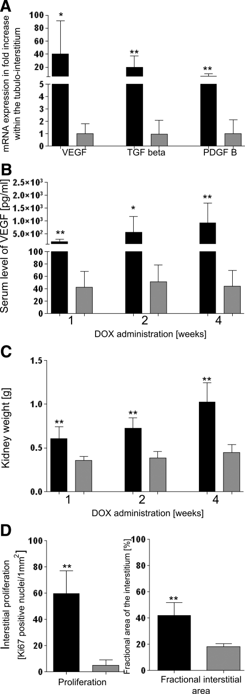
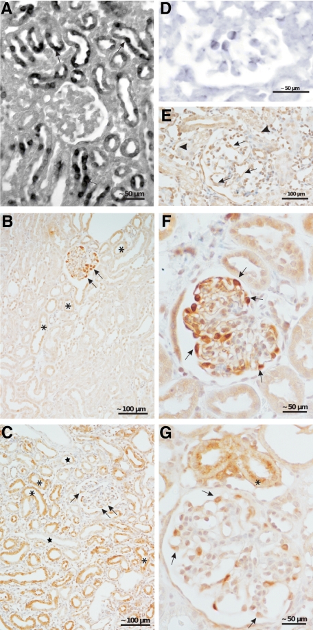
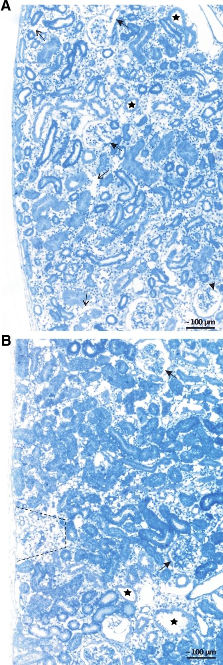
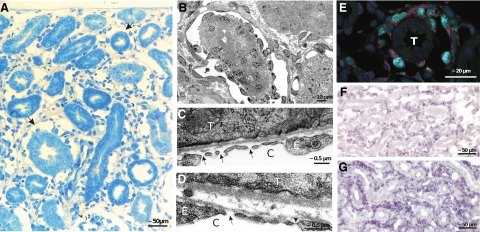
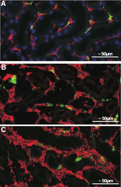
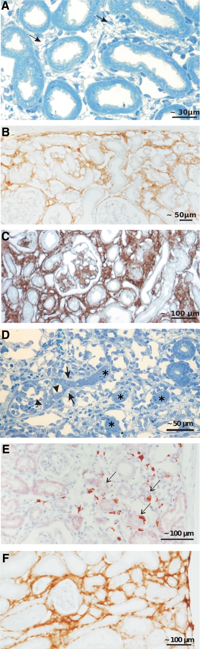
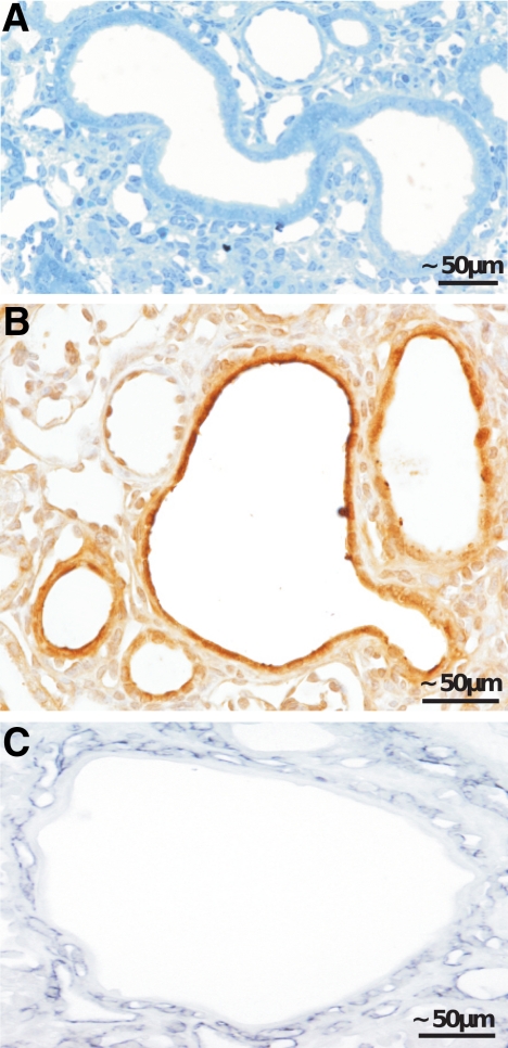
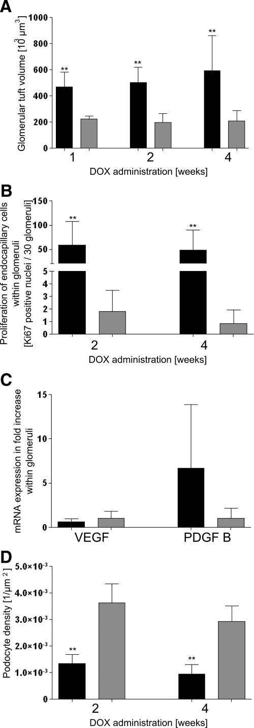
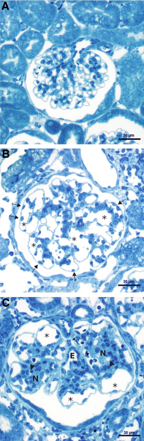
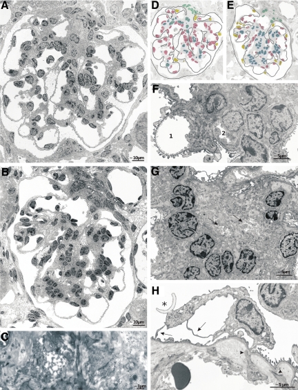
The role of vascular endothelial growth factor (VEGF) in renal fibrosis, tubular cyst formation, and glomerular diseases is incompletely understood. We studied a new conditional transgenic mouse system [Pax8-rtTA/(tetO)(7)VEGF], which allows increased tubular VEGF production in adult mice. The following pathology was observed. The interstitial changes consisted of a ubiquitous proliferation of peritubular capillaries and fibroblasts, followed by deposition of matrix leading to a unique kind of fibrosis, ie, healthy tubules amid a capillary-rich dense fibrotic tissue. In tubular segments with high expression of VEGF, cysts developed that were surrounded by a dense network of peritubular capillaries. The glomerular effects consisted of a proliferative enlargement of glomerular capillaries, followed by mesangial proliferation. This resulted in enlarged glomeruli with loss of the characteristic lobular structure. Capillaries became randomly embedded into mesangial nodules, losing their filtration surface. Serum VEGF levels were increased, whereas endogenous VEGF production by podocytes was down-regulated. Taken together, this study shows that systemic VEGF interferes with the intraglomerular cross-talk between podocytes and the endocapillary compartment. It suppresses VEGF secretion by podocytes but cannot compensate for the deficit. VEGF from podocytes induces a directional effect, attracting the capillaries to the lobular surface, a relevant mechanism to optimize filtration surface. Systemic VEGF lacks this effect, leading to severe deterioration in glomerular architecture, similar to that seen in diabetic nephropathy.


Figures

Figure 1
Figure 1
Values of experimental animals in black, controls in gray columns; shown are the means ± SD. A: mRNA expression within the tubulo-interstitium after 4 weeks of DOX; compared with controls, expression of VEGF, TGF-β, and PDGF-B is up-regulated. N (transgenics) = 10 (tubulo-interstial and total kidney samples were pooled), N (controls) = 9; , P < 0.05; , P < 0.01. B: Serum levels of VEGF. After 1, 2, and 4 weeks of DOX, transgenic animals exhibit increased serum levels of VEGF in comparison with control mice. N = 7; , P < 0.05; , P < 0.01. C: Kidney weight. After 1, 2, and 4 weeks of DOX, a dramatic increase in kidney weight is noticed in transgenic mice compared with controls. N = 7; , P < 0.01. D: Interstitial cell proliferation and fractional interstitial area. After 2 weeks of DOX, transgenic animals show a strongly elevated number of Ki-67-positive cells within the peritubular interstitium (comprising all interstitial cells including endothelial cells) together with an enormous expansion of the peritubular interstitium. N = 7; , P < 0.01.
Figure 2
Figure 2
VEGF and PDGF-B. A: In situ hybridization of VEGF. In transgenic animals a heterogeneous tubular expression of VEGF mRNA (arrows) is encountered; no signal is seen with the sense probe (data not shown). B, C, F, and G: Immunohistochemistry of VEGF. B and F: In controls, a strong staining in podocytes (arrows) and a very weak staining in some tubules (asterisks) are seen. C and G: In transgenic animals, a strong but heterogenous staining is encountered; the majority of tubules shows a strong staining (asterisk); a couple of tubules (stars) do not show any reaction. Staining intensity in podocytes is decreased (arrows). D: In situ hybridization of PDGF-B. Note the expression in glomerular endothelial cells. In controls, no reaction was seen (data not shown). E: Immunohistochemistry reveals a strong expression PDGF-B in glomerular (arrows) and peritubular endothelial cells (arrowheads). Two weeks of DOX (A, D, and E); 4 weeks of DOX (B and C).
Figure 3
Figure 3
Renal cortex; overviews. A: After 2 weeks of DOX, ubiquitous expansion of the peritubular space with an increased density of interstitial cells and enlargement of capillaries (thin arrows). Tubules are heterogeneous in diameter; some of them (stars) show the beginning of cyst formation. Glomeruli show enlarged afferent arterioles (arrows), dilated capillaries (arrowhead), and focal centers of mesangial expansion. B: After 4 weeks, the expansion of the peritubular spaces is largely preserved; tubules in such areas look normal. In circumscribed areas (marked by a hatched line), tubules undergo atrophy/degeneration corresponding to the area of a nephron in which degeneration started from a glomerular damage. Individual tubular cysts (stars) are also encountered. Glomeruli show nodule formation (arrows).
Figure 4
Figure 4
Peritubular interstitium and capillaries after 2 weeks of DOX. A: Within the expanded interstitium capillaries are enlarged at many sites to giant vascular channels completely surrounding a tubular profile (arrows). Note the abundance of interstitial cells and matrix; tubules are normal. B: Enlarged peritubular capillary completely surrounding a tubular profile; note the high density of endothelial cells. Frequently, areas equipped with a porous endothelium, ie, with undiaphragmed fenestrae are observed (arrows in C and D); the arrowhead in D points to a diaphragmed fenestra for comparison. C, capillary; E, endothelium; T, tubule. E: Immunofluorescence. Double-labeling against proliferating cell nuclear antigen (green) and CD31 (red) emphasizes the vivid proliferation of endothelial cells of an enlarged peritubular capillary; T, tubule. F and G: In situ hybridization reveals a prominent expression of PDGF-B (F) and TGF-β (G) in cells of the peritubular interstitium, mostly probably endothelial cells. In controls, no signals were observed; sense probes were negative (data not shown).
Figure 5
Figure 5
Expanded peritubular spaces after 2 weeks of DOX. A–C: Immunofluorescence. Staining against 5′NT (red in A and B) and α-SMA (red in C) and major histocompatibility complex class II (green). A pronounced increase of 5′NT-positive fibroblasts is seen in B compared with the control (A). A dramatic appearance of α-SMA-positive myofibroblasts is seen in C; in controls, no myofibroblasts are observed (data not shown). No differences in major histocompatibility complex class II-positive cells (green) are seen.
Figure 6
Figure 6
Changes within the interstitium. A–C: Development of fibrosis around intact tubules. Already after 2 weeks of DOX (A), more pronounced after 4 weeks (C and D), a dense fibrotic tissue (arrows in A) is encountered that contains collagen I (B) and collagen IV (C) as shown by immunohistochemistry. D–F: Focal interstitial fibrosis associated with the atrophy/degeneration of a nephron. D: Glomerular profile with tuft adhesion and proliferative process at Bowman’s capsule (between two arrowheads), expansion of the parietal basement membrane (arrows), and encroachment on the tubule. The complete obstruction of the urinary orifice was verified by tracing in serial sections. Note the corresponding atrophic tubules (asterisks) in the vicinity surrounded by fibrous tissue. E and F: Immunohistochemistry showing the numerous macrophages (E; ER-HR3 staining) and the dense accumulation of collagen I in such areas (F). Arrows in E denote red-stained macrophages.
Figure 7
Figure 7
Cyst formation within renal tubules. A–C: Tubular cysts of a proximal tubule (A). B and C: Immunohistochemistry showing the strong staining against VEGF in the cystic epithelium (B) and the high density of capillaries surrounding the cyst (C; shown with an antibody against Meca 32).
Figure 8
Figure 8
Values of experimental animals in black, controls in gray columns; shown are the means ± SD. A: Glomerular tuft volume; a dramatic increase is seen after 1, 2, and 4 weeks of DOX in transgenic animals. N = 7; P < 0.01. B: Proliferation of endocapillary cells within glomeruli. After 2 and 4 weeks of DOX, the number of Ki-67-positive endocapillary cells (including mesangial and endothelial cells) is drastically increased in transgenic mice. N = 7; P < 0.01. C: mRNA expression within glomeruli. After 4 weeks of DOX, VEGF within glomeruli of transgenic animals shows a tendency to decrease whereas PDGF-B increases. N = 4. D: Podocyte density. Transgenic animals show a dramatic decrease in podocyte density of glomeruli after 2 and 4 weeks of DOX. N (transgenics) = 7, N (controls) = 5; P < 0.01.
Figure 9
Figure 9
Glomerular changes. A: Control; note the numerous uniformly small capillary profiles. B: After 2 weeks of DOX, giant glomeruli with fewer but considerably lager capillary profiles (asterisks) are prominent. Frequently, endothelial cell bodies are lying at unusual peripheral positions (arrows). Note the islands of clustered mesangium. C: After 4 weeks of DOX, individual giant glomerular capillaries (asterisks) are still seen, otherwise mesangial nodules (N) developed that contain either small or completely collapsed capillaries (arrowheads). Note the intraglomerular segment of the efferent arteriole (E) that is densely equipped with endothelial cells (seen in both glomeruli).
Figure 10
Figure 10
Glomerular changes; TEM details. A, B, D, and E: Glomerular profiles after 2 (A and D) and 4 weeks (B and E) of DOX treatment; D and E indicate the cellular composition in both profiles (nuclei of endothelial cells are shown in red, of mesangial cells in blue, of podocytes in yellow, and of extraglomerular mesangial cells in turquoise). After 2 weeks of DOX capillary enlargement and endothelial proliferation (red-labeled cells) and after 4 weeks mesangial proliferation (blue-labeled cells) are dominant. Note that the podocyte foot processes are generally well preserved. C: A capillary in the process of strangulation and collapse. F: A stage after 4 weeks of DOX showing the incorporation of capillaries (1 and 2) into an expanding mesangial nodule. Mesangial cells extend toward the periphery at both sides of 1 and the almost total embracement and collapse of 2; note that the podocyte foot processes of this capillary are partially effaced. G: A mesangial nodule consisting of cells and matrix that contains the collapsed remnants of at least two capillaries (arrows). H: An apparently hypoplastic podocyte with primary processes (arrow) that are extended into thin sheets. Note the partial foot process effacement and the shedding of a portion of an apical membrane (asterisk) into Bowman’s space. Furthermore, the separation of the endothelium from the GBM by the expanding mesangium is seen; mesangial cell processes still fix to the GBM (arrowheads).

References

    1. Holmes DI, Zachary I. The vascular endothelial growth factor (VEGF) family: angiogenic factors in health and disease. Genome Biol. 2005;6:209. - PMC - PubMed
    1. Kriz W. Ontogenetic development of the filtration barrier. Nephron Exp Nephrol. 2007;106:e44–e50. - PubMed
    1. Eremina V, Quaggin SE. The role of VEGF-A in glomerular development and function. Curr Opin Nephrol Hypertens. 2004;13:9–15. - PubMed
    1. Eremina V, Sood M, Haigh J, Nagy A, Lajoie G, Ferrara N, Gerber HP, Kikkawa Y, Miner JH, Quaggin SE. Glomerular-specific alterations of VEGF-A expression lead to distinct congenital and acquired renal diseases. J Clin Invest. 2003;111:707–716. - PMC - PubMed
    1. Schrijvers BF, Flyvbjerg A, De Vriese AS. The role of vascular endothelial growth factor (VEGF) in renal pathophysiology. Kidney Int. 2004;65:2003–2017. - PubMed

Publication types

MeSH terms

Substances