

Fundus autofluorescence patterns in eyes with primary intraocular lymphoma
- PMID: 19834356
- PMCID: PMC3707141
- DOI: 10.1097/IAE.0b013e3181b408a2
Fundus autofluorescence patterns in eyes with primary intraocular lymphoma
Abstract
Purpose: To evaluate the fundus autofluorescence (FAF) patterns in the eyes with primary intraocular lymphomas (PIOLs).
Methods: A review of the medical charts of four consecutive patients (five eyes) with PIOL who had been studied by FAF. A fundus camera was used to obtain the FAF images. Optical coherence tomography was also performed.
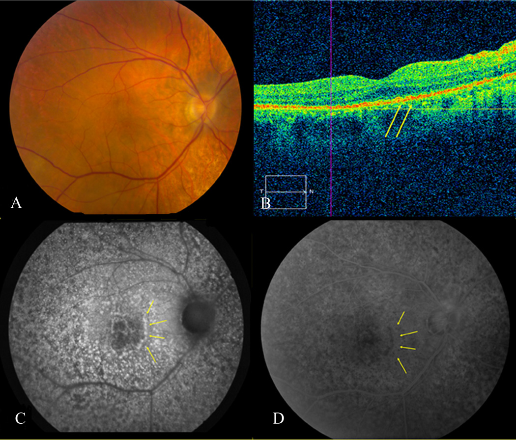
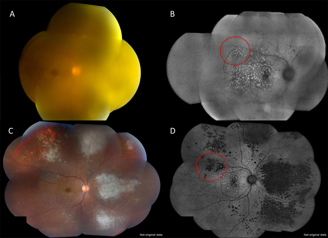
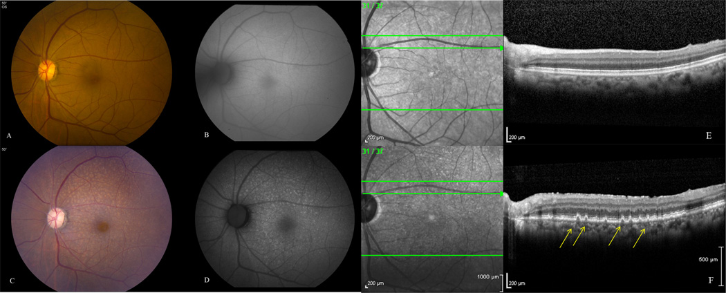
Results: The ophthalmoscopically observed brown clumps on the surface of greasy yellowish masses beneath the retinal pigmented epithelium had a bright hyperfluorescence appearance by FAF. This FAF hyperfluorescence was completely reversed to hypofluorescence in the fluorescein angiograms. The diffuse infiltration of the cells making up the PIOL above the retinal pigmented epithelium was ophthalmoscopically observed as a retinal whitening and was hypofluorescent by FAF. These areas of hypofluorescence were also reversed to areas of hyperfluorescence in the fluorescein angiogram. Fundus autofluorescence clearly delineated the retinal pigmented epithelium atrophy, which developed after the spontaneous resolution of the PIOL as a hypoautofluorescent area.
Conclusion: Because FAF can reveal various findings of PIOLs, it can be used to differentiate the patients with PIOL from those with ocular inflammatory diseases. Although further studies are required to determine whether these findings are characteristic to PIOL, this noninvasive method can then lead to earlier diagnosis and treatment.
Conflict of interest statement
None of the authors have any conflict of interest in the materials presented in this manuscript.
Figures
References
-
- Coupland SE, Damato B. Understanding intraocular lymphomas. Clinical and Experimental Ophthalmology. 2008;36(6):564–578. - PubMed
-
- Grimm SA, Pulido JS, Jahnke K, et al. Primary intraocular lymphoma: an international primary central nervous system lymphoma collaborative group report. Annals of Oncology. 2007;18(11):1851–1855. - PubMed
-
- Choi JY, Kafkala C, Foster CS. Primary intraocular lymphoma: A review. Semin Ophthalmol. 2006 Jul-Sep;21(3):125–133. Review. - PubMed
Publication types
MeSH terms
Substances
Grants and funding
LinkOut - more resources
Full Text Sources
