

Comparison of fat-water MRI and single-voxel MRS in the assessment of hepatic and pancreatic fat fractions in humans
- PMID: 19834463
- PMCID: PMC2847037
- DOI: 10.1038/oby.2009.352
Comparison of fat-water MRI and single-voxel MRS in the assessment of hepatic and pancreatic fat fractions in humans
Abstract
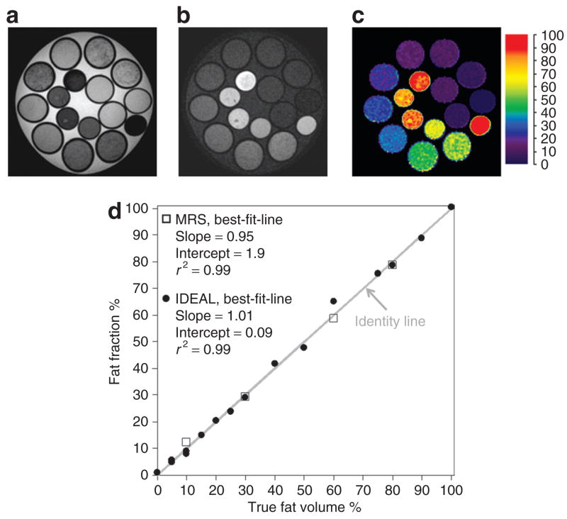
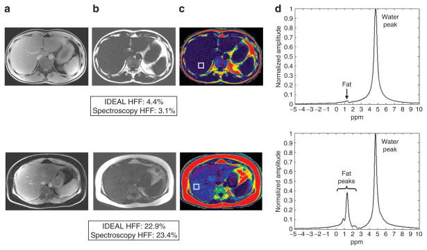
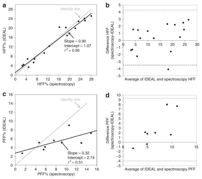
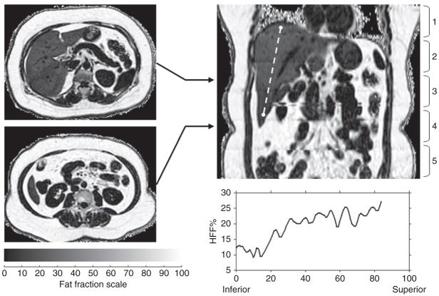
The ability to accurately and noninvasively quantify fatty infiltration in organs such as the liver and the pancreas remains a critical component in understanding the link between obesity and its comorbidities such as type 2 diabetes and fatty liver disease. Single-voxel ((1)H) proton magnetic resonance spectroscopy (MRS) has long been regarded as the gold-standard noninvasive technique for such measurements. Recent advances in three-dimensional fat-water magnetic resonance imaging (MRI) methods have led to the development of rapid, robust, and quantitative approaches that can accurately characterize the proportion of fat and water content in underlying tissues across the full imaging volume, and hence entire organs of interest. One such technique is called IDEAL (Iterative Decomposition with Echo Asymmetry and Least squares estimation). This article prospectively compares three-dimensional (3D) IDEAL-MRI and single-voxel MRS in the assessment of hepatic (HFF) and pancreatic fat fraction (PFF) in 16 healthy subjects. MRS acquisitions took 3-4 min to complete whereas IDEAL acquisitions were completed in 20-s breath-holds. In the liver, there was a strong correlation (slope = 0.90, r(2) = 0.95, P < 0.001) between IDEAL and MRS-based fat fractions. In the pancreas, a poorer agreement between IDEAL and MRS was observed (slope = 0.32, r(2) = 0.51, P < 0.02). The discrepancy of PFF is likely explained by MRS signal contamination from surrounding visceral fat, presumably during respiratory motion. We conclude that IDEAL is equally accurate in characterizing hepatic fat content as MRS, and is potentially better suited for fat quantification in smaller organs such as the pancreas.
Conflict of interest statement
The authors declared no conflict of interest.
Figures
References
-
- Ogden CL, Carroll MD, Curtin LR, et al. Prevalence of overweight and obesity in the United States, 1999–2004. JAMA. 2006;295:1549–1555. - PubMed
-
- Després JP. Cardiovascular disease under the influence of excess visceral fat. Crit Pathw Cardiol. 2007;6:51–59. - PubMed
-
- Bergman RN, Kim SP, Hsu IR, et al. Abdominal obesity: role in the pathophysiology of metabolic disease and cardiovascular risk. Am J Med. 2007;120:S3–S8. discussion S29. - PubMed
-
- Després JP, Lemieux I, Bergeron J, et al. Abdominal obesity and the metabolic syndrome: contribution to global cardiometabolic risk. Arterioscler Thromb Vasc Biol. 2008;28:1039–1049. - PubMed
-
- Mathieu P, Pibarot P, Larose E, et al. Visceral obesity and the heart. Int J Biochem Cell Biol. 2008;40:821–836. - PubMed
Publication types
MeSH terms
Substances
Grants and funding
LinkOut - more resources
Full Text Sources
Other Literature Sources
Medical
