. 2009;4(11):1614-22.
doi: 10.1038/nprot.2009.186. Epub 2009 Oct 15.

Differentiation of human oligodendrocytes from pluripotent stem cells

Affiliations

Differentiation of human oligodendrocytes from pluripotent stem cells

Bao-Yang Hu et al. Nat Protoc. 2009.

Abstract

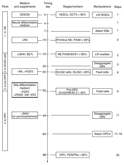
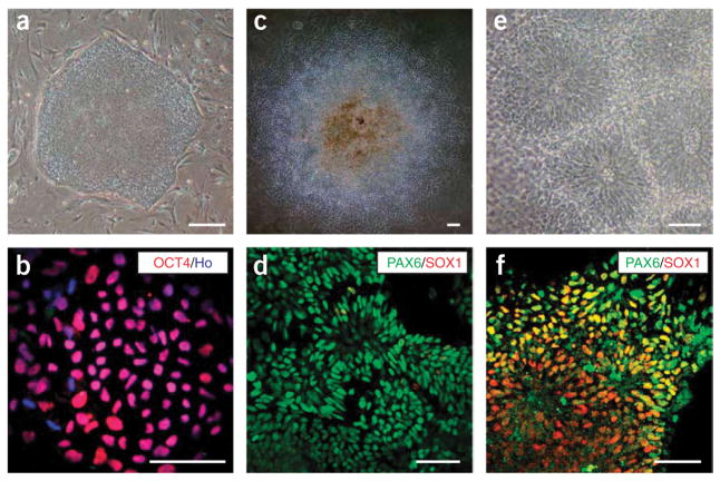
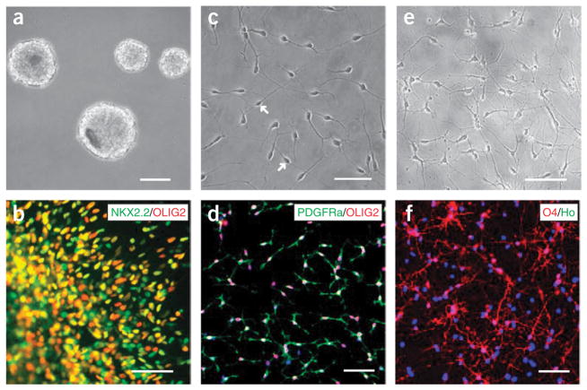
We have developed a four-part protocol to differentiate human embryonic stem cells (hESCs) to oligodendrocyte progenitor cells (OPCs) according to developmental principles. In the first 2 weeks, hESCs are induced to differentiate into neuroepithelial cells, which form neural tube-like rosettes. In the following 10 d, these neuroepithelial cells are specified to OLIG2-expressing progenitors in the presence of retinoic acid (RA) and sonic hedgehog (SHH). Upon treatment with fibroblast growth factor 2 (FGF2) for another 10 d, these progenitors convert to OLIG2 and NKX2.2-expressing pre-OPCs. Finally, the pre-OPCs take 8-9 weeks to differentiate into OPCs, which express additional markers of oligodendrocytes, such as SOX10, platelet-derived growth factor receptor alpha (PDGFRalpha) and NG2. The unique aspects of the protocol are the use of FGF2 to promote the differentiation of gliogenic pre-OPCs in the third part and the removal of FGF2 during the transition of pre-OPCs to OPCs. This 3-month differentiation protocol consistently yields OPCs of high purity capable of producing myelin sheaths in vivo.


Figures

Figure 1
Figure 1
Flowchart of the four-part oligodendrocyte progenitor cell (OPC) differentiation from human embryonic stem cells (hESCs). Part one differentiates the hESCs to PAX6+ neuroepithelial cells in the serum free medium without morphogens for 2 weeks. Part two patterns the neureopithelia to OLIG2+ ventral progenitors by retinoic acid (RA) and sonic hedgehog (SHH) (or purmorphamine). In part three, the neurogenic potential of the OLIG2 progenitors is repressed by removal of RA and addition of fibroblast growth factor 2 (FGF2) for 10 d. The OLIG2 progenitors now co-express NKX2.2 and become gliogenic, which we refer to as pre-OPCs. Finally, the pre-OPCs are differentiated to OPCs in the glia differentiation medium, which lacks FGF2, and contains platelet-derived growth factor (PDGF), insulin like growth factor (IGF) and neurotrophin 3 (NT3). This fourth part takes 8–9 weeks. The OPCs express SOX10, platelet-derived growth factor receptor alpha (PDGFRα), NG2 in addition to OLIG2 and NKX2.2. This chart is modified from Figure 1 of our motoneuron differentiation protocol. hESCM, human embryonic stem cell medium; IGF, insulin like growth factor; NE, neuroepithelial cells; PDGF, platelet derived growth factor; NT3, neurotrophin 3. Percentage denotes the promotion of positive cells among total progenies from the starting hESCs.
Figure 2
Figure 2
Induction of neuroepithelial cells. Human embryonic stem cells (hESCs) growing on mouse embryonic fibroblast (MEF) feeder cells as a uniform colony (a). All cells are positively stained for OCT4 (b). On day 10, columnar epithelial cells appear and organize into rosettes in the colony (c). These cells express PAX6 but not SOX1 (d). At day 15, neural tube-like rosettes are obvious (e) and cells within these rosettes are stained for both PAX6 and SOX1 (f). Scale bar, 50 μm. Ho: Hoechst 33258.
Figure 3
Figure 3
Generation of pre-oligodendrocyte progenitor cells (OPCs) and OPCs. The pre-OPCs grow as spheres in suspension at day 35 (a) and express both OLIG2 and NKX2.2 (b). Dissociated OPCs after 14 weeks of human embryonic stem cell (hESC) differentiation exhibit a bipolar morphology (c, arrows), and co-express OLIG2 and platelet-derived growth factor receptor alpha (PDGFRα) (d). At 4 weeks after growing on substrate for differentiation, OPCs become multipolar immature oligodendrocytes (e), and many of these cells express 04 (f). Scale bar, 50 μm. Ho denotes Hoechst 33258.

References

    1. Lu QR, et al. Sonic hedgehog-regulated oligodendrocyte lineage genes encoding bHLH proteins in the mammalian central nervous system. Neuron. 2000;25:317–329. - PubMed
    1. Zhou Q, Wang S, Anderson DJ. Identification of a novel family of oligodendrocyte lineage-specific basic helix-loop-helix transcription factors. Neuron. 2000;25:331–343. - PubMed
    1. Sugimori M, et al. Combinatorial actions of patterning and HLH transcription factors in the spatiotemporal control of neurogenesis and gliogenesis in the developing spinal cord. Development. 2007;134:1617–1629. - PubMed
    1. Lee SK, Lee B, Ruiz EC, Pfaff SL. Olig2 and Ngn2 function in opposition to modulate gene expression in motor neuron progenitor cells. Genes Dev. 2005;19:282–294. - PMC - PubMed
    1. Marquardt T, Pfaff SL. Cracking the transcriptional code for cell specification in the neural tube. Cell. 2001;106:651–654. - PubMed

Publication types

MeSH terms