. 2009 Oct;5(10):e1000683.
doi: 10.1371/journal.pgen.1000683. Epub 2009 Oct 16.

Limiting the persistence of a chromosome break diminishes its mutagenic potential

Affiliations

Limiting the persistence of a chromosome break diminishes its mutagenic potential

Nicole Bennardo et al. PLoS Genet. 2009 Oct.

Abstract

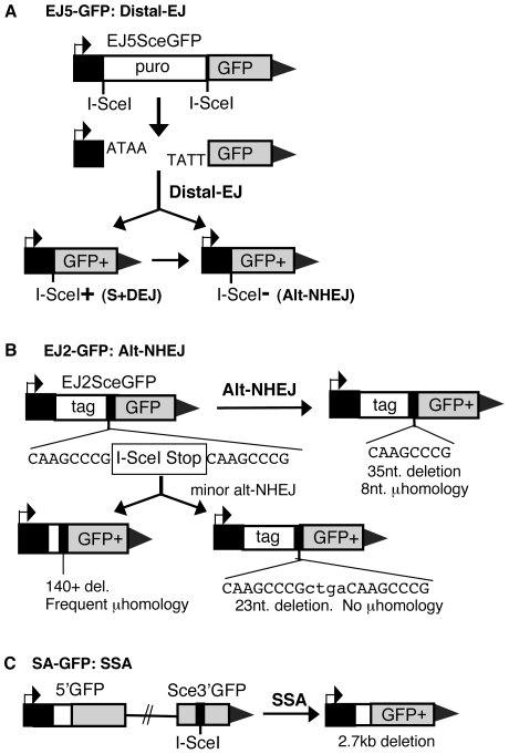
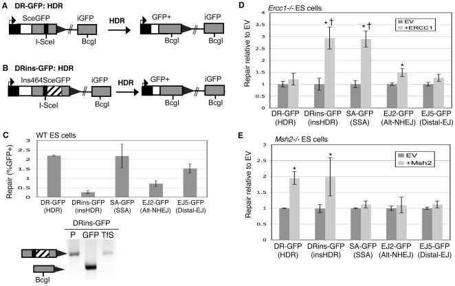
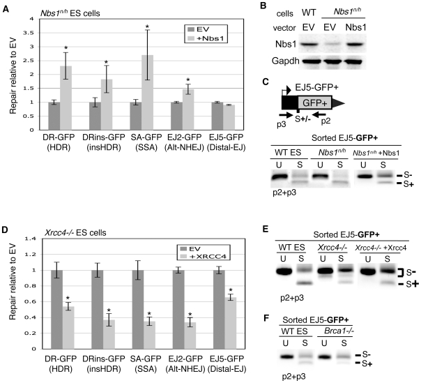
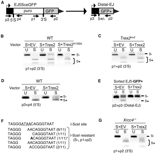
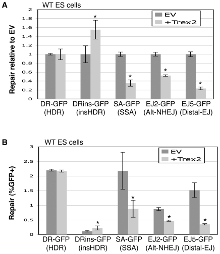
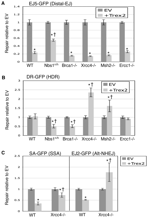
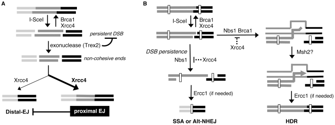
To characterize the repair pathways of chromosome double-strand breaks (DSBs), one approach involves monitoring the repair of site-specific DSBs generated by rare-cutting endonucleases, such as I-SceI. Using this method, we first describe the roles of Ercc1, Msh2, Nbs1, Xrcc4, and Brca1 in a set of distinct repair events. Subsequently, we considered that the outcome of such assays could be influenced by the persistent nature of I-SceI-induced DSBs, in that end-joining (EJ) products that restore the I-SceI site are prone to repeated cutting. To address this aspect of repair, we modified I-SceI-induced DSBs by co-expressing I-SceI with a non-processive 3' exonuclease, Trex2, which we predicted would cause partial degradation of I-SceI 3' overhangs. We find that Trex2 expression facilitates the formation of I-SceI-resistant EJ products, which reduces the potential for repeated cutting by I-SceI and, hence, limits the persistence of I-SceI-induced DSBs. Using this approach, we find that Trex2 expression causes a significant reduction in the frequency of repair pathways that result in substantial deletion mutations: EJ between distal ends of two tandem DSBs, single-strand annealing, and alternative-NHEJ. In contrast, Trex2 expression does not inhibit homology-directed repair. These results indicate that limiting the persistence of a DSB causes a reduction in the frequency of repair pathways that lead to significant genetic loss. Furthermore, we find that individual genetic factors play distinct roles during repair of non-cohesive DSB ends that are generated via co-expression of I-SceI with Trex2.


Conflict of interest statement

The authors have declared that no competing interests exist.

Figures

Figure 1
Figure 1. Reporters for EJ and SSA repair pathways.
(A) EJ5-GFP is shown along with products of EJ between distal DSB ends (Distal-EJ) that restores a GFP expression cassette, including Ku-dependent I-SceI site restoration (S+DEJ), and Ku-independent I-SceI-resistant products (Alt-NHEJ). (B) EJ2-GFP is shown along with products of Alt-NHEJ that restore the reading frame of the GFP expression cassette. The most abundant Alt-NHEJ product involves 8 nt of microhomology at the repair junction and a 35 nt deletion. (C) SA-GFP is shown along with the SSA repair product that utilizes 266 nt of homology between the tandem GFP segments, thereby restoring a GFP expression cassette.
Figure 2
Figure 2. Ercc1 promotes repair pathways that require removal of a nonhomologous segment, whereas Msh2 promotes HDR.
(A) DR-GFP is shown along with the HDR product that uses iGFP as the template for nascent DNA synthesis, which results in restoration of a GFP expression cassette. (B) Shown is a distinct reporter for HDR (DRins-GFP) that is similar to DR-GFP, except it contains an insertion of 464 nt downstream of the I-SceI site that needs to be removed during HDR to restore a GFP expression cassette. (C) Requiring removal of a nonhomologous insertion during HDR decreases the efficiency of repair. DR-GFP and DRins-GFP were integrated at the identical locus (Pim1) of wild-type (J1 strain) mouse ES cells, and the efficiency of HDR (%GFP+ cells) was determined following transient expression of I-SceI. Shown is the efficiency of repair (%GFP+ cells) from these reporters, along with those from Figure 1 that are also in WT ES cells. Also shown is confirmation of the structure of the GFP+ repair product for DRins-GFP via PCR/sequencing analysis of sorted GFP+ cells. The labels P, GFP, and TfS denote the parental cell line, GFP+ sorted cells, and total I-SceI-transfected cells, respectively. (D) Ercc1 specifically promotes DSB repair pathways that require removal of an extended nonhomologous segment. Each of the reporters depicted in Figure 1 and Figure 2A and 2B were integrated into mouse ES cells deficient in Ercc1. These individual cell lines were transfected with an expression vector for I-SceI, along with either a complementation vector for Ercc1, or the empty expression vector (EV). Repair is measured as percent GFP+ cells, which is normalized to the EV samples transfected in parallel. Asterisks denote a statistical difference in repair efficiency from EV (p<0.0001). The dagger denotes a statistical difference in fold-complementation compared to HDR of the DR-GFP reporter (p<0.0001). (E) Msh2 specifically promotes HDR. Analysis of repair in Msh2−/− cells was performed as described above for Ercc1, where asterisks denote the same statistical differences as described above.
Figure 3
Figure 3. Nbs1, Xrcc4, and Brca1 play distinct roles during individual repair events.
(A) Nbs1 promotes HDR, SSA, and Alt-NHEJ, but is dispensable for Distal-EJ. Reporters from Figure 1 and Figure 2 were integrated into Nbs1n/h cells, and the effect of Nbs1 complementation on repair was determined as for Ercc1 in Figure 2D. Asterisks denote a statistical difference in repair efficiency from EV (p<0.0001). (B) Nbs1n/h cells show a reduced level of Nbs1 that is restored to wild-type levels with transient expression. Shown are immunoblot signals for Nbs1 from transfections with Nbs1n/h, and from WT cells. (C) The repair event S+DEJ is increased in Nbs1-deficient cells. The diagram depicts the primers used for amplification (p2, p3). Shown are amplification products from sorted GFP+ cells derived from I-SceI transfection of EJ5-GFP-containing WT and Nbs1n/h cells, as well as Nbs1n/h cells transiently complemented with Nbs1. The products have been left uncut (U) and cut with I-SceI (S). (D) Xrcc4 suppresses HDR, SSA, Alt-NHEJ, and Distal-EJ. Shown are repair levels of reporters integrated into Xrcc4−/− ES cells that were assayed with/without transient complementation of Xrcc4 as described for Ercc1 in Figure 2D. Asterisks denote a statistical difference in repair efficiency from EV (p<0.0001). (E) Xrcc4-deficient cells show a decrease in S+DEJ. Shown are amplification products using the same primers and annotation as shown in C, from sorted GFP+ cells derived from I-SceI transfection of EJ5-GFP-containing WT cells, Xrcc4−/− cells, and Xrcc4−/− cells transiently complemented with Xrcc4 as in D. (F) Brca1-deficient cells show a decrease in S+DEJ. As in E, amplification products are shown from sorted GFP+ cells derived from I-SceI transfection of EJ5-GFP for WT and Brca1−/− cell lines.
Figure 4
Figure 4. Expression of the Trex2 exonuclease promotes the formation of Xrcc4-dependent I-SceI-resistant EJ products.
(A) Shown are primers for EJ5-GFP that are used to analyze proximal-EJ at the 5′ and 3′ I-SceI sites (shown as 5′S and 3′S, respectively), as well as Distal-EJ. (B) Expression of the Trex2 exonuclease promotes the formation of I-SceI-resistant EJ products between proximal DSB ends at the 3′ I-SceI site in EJ5-GFP. WT ES cells with EJ5-GFP were transfected with an I-SceI expression vector (S) along with a Trex2 expression vector (Trex2), an exonuclease-deficient mutant of Trex2 (Trex2-H188A), or EV. Shown are amplification products from these transfections using the primers p1 and p2, which were either left uncut (U) or were cut with I-SceI (S). (C) Trex2 expression also promotes I-SceI-resistant EJ products in Trex2null cells. Analysis was performed on Trex2null cells with the EJ5-GFP reporter as described in B. (D) Trex2 expression promotes I-SceI-resistant EJ products at the 5′ I-SceI site of EJ5-GFP. Shown are amplification products using the primers p3 and p4, using the same transfection conditions and annotation as described in B. (E) Trex2 expression promotes I-SceI-resistant Distal-EJ products. Shown are amplification products from sorted GFP+ cells derived from the transfections shown in C, using primers p3 and p2, with the same annotation shown in B. (F) EJ products via Trex2 show deletion of segments of the I-SceI 3′ overhang (underlined). The I-SceI-resistant products shown in C were cloned, and 11 individual clones were sequenced. Shown are the sequences of these clones, where the numerator in parenthesis depicts the number of times a given sequence was identified. An asterisk denotes the one clone with evidence of microhomology (1 nt., A in bold). (G) I-SceI-resistant proximal EJ products via Trex2 are dependent on Xrcc4. Analysis of the effect on Trex2 expression on the 3′ I-SceI site of the EJ5-GFP reporter was performed on Xrcc4−/− cells as described in B.
Figure 5
Figure 5. Trex2 expression causes a significant decrease in Distal-EJ, Alt-NHEJ, and SSA, but not HDR.
WT ES cell lines with individual reporters were co-transfected with expression vectors for I-SceI and Trex2. (A) Shown are repair values normalized to parallel co-transfections with I-SceI and EV. Asterisks denote a statistical difference from EV (p<0.0001, DRins-GFP p = 0.0008). (B) Shown are primary repair values (%GFP+) from the experiment shown in A, to allow a direct comparison of the frequencies of different repair events. The error bars are somewhat larger in this panel as compared to A, since the primary repair levels show greater experimental variation versus the relatively consistent fold-effect of Trex2 expression. Asterisks denote a statistical difference from EV (p<0.0001 for EJ2-GFP and EJ5-GFP, p = 0.001 for SA-GFP, and p = 0.005 for DRins-GFP).
Figure 6
Figure 6. Roles of individual genetic factors during repair of DSBs with non-cohesive ends generated by co-expression of I-SceI with Trex2.
Cell lines with individual reporters were co-transfected with expression vectors for I-SceI and Trex2. Repair values are quantified and normalized to parallel co-transfections with I-SceI and EV. (A) Nbs1-deficient cells show a diminished suppression of Distal-EJ from Trex2 expression. Shown are the effects of Trex2 expression on the EJ5-GFP reporter integrated into DNA repair-deficient mouse ES cell lines described in Figure 2 and Figure 3. Asterisks denote a statistical difference from EV (p<0.0001), and the dagger denotes a statistical difference from WT (p<0.0001). (B) Trex2 expression causes a decrease in HDR in cells deficient in Nbs1 and Brca1, but an increase in HDR in cells deficient in Xrcc4 and Msh2. Shown are the effects of Trex2 expression on the DR-GFP reporter in the cell lines shown in A. Asterisks denote a statistical difference from EV (p<0.0001, Msh2 p = 0.001), and the dagger denotes a statistical difference from WT (p<0.0001, Msh2 p = 0.0027). (C) Trex2 expression in Xrcc4−/− cells shows an increase in Alt-NHEJ, and a relative increase in SSA compared to WT. Shown are the effects of Trex2 expression on the EJ2-GFP and SA-GFP reporters in Xrcc4−/− cells, along with WT. Asterisks and daggers are as in A.
Figure 7
Figure 7. Limiting the persistence of a DSB causes a reduction in repair pathways that result in genetic loss.
(A) Proximal EJ is shown to limit Distal-EJ of two tandem DSBs. Xrcc4 and Brca1 are shown to promote EJ of cohesive DSB ends, where repeated cutting by I-SceI results in a persistent DSB. The Trex2 exonuclease is shown as generating non-cohesive DSB ends, which limits the persistence of I-SceI-induced DSBs. Xrcc4 is also shown promoting EJ of non-cohesive ends during both proximal-EJ and Distal-EJ. The bias towards proximal-EJ in WT cells is indicated by the bold arrow (see Figure 5). Similarly, the moderate preference for proximal-EJ via Xrcc4 is indicated by the bold-type. (B) Shown is a model whereby the persistent nature of a DSB is important for SSA and Alt-NHEJ, but not HDR. Nbs1 and Brca1 are shown having an increased role during HDR of a relatively less persistent DSB. End resection that leads to HDR, SSA, and Alt-NHEJ is shown as being suppressed by Xrcc4 and promoted by Nbs1. Msh2 is shown as possibly promoting 3′ end processing prior to nascent DNA synthesis during HDR. Ercc1 is shown to promote the completion of repair pathways if needed, based on the requirement for removal of an extended nonhomologous segment.

References

    1. Wyman C, Kanaar R. DNA double-strand break repair: all's well that ends well. Annu Rev Genet. 2006;40:363–383. - PubMed
    1. Greaves MF, Wiemels J. Origins of chromosome translocations in childhood leukaemia. Nat Rev Cancer. 2003;3:639–649. - PubMed
    1. Ma Y, Lu H, Schwarz K, Lieber MR. Repair of double-strand DNA breaks by the human nonhomologous DNA end joining pathway: the iterative processing model. Cell Cycle. 2005;4:1193–1200. - PubMed
    1. Haber JE. Alternative endings. Proceedings of the National Academy of Sciences. 2008;105:405–406. - PMC - PubMed
    1. McVey M, Lee SE. MMEJ repair of double-strand breaks (director's cut): deleted sequences and alternative endings. Trends in Genetics. 2008;24:529–538. - PMC - PubMed

Publication types

MeSH terms

Substances