. 2009 Oct 16;4(10):e7485.
doi: 10.1371/journal.pone.0007485.

Sequence and structure signatures of cancer mutation hotspots in protein kinases

Affiliations

Sequence and structure signatures of cancer mutation hotspots in protein kinases

Anshuman Dixit et al. PLoS One. .

Abstract

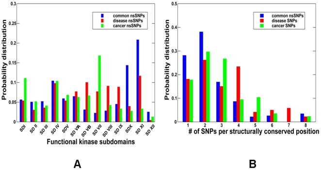
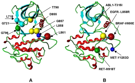
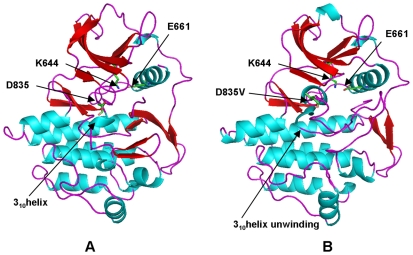
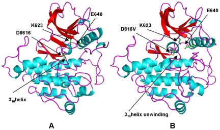
Protein kinases are the most common protein domains implicated in cancer, where somatically acquired mutations are known to be functionally linked to a variety of cancers. Resequencing studies of protein kinase coding regions have emphasized the importance of sequence and structure determinants of cancer-causing kinase mutations in understanding of the mutation-dependent activation process. We have developed an integrated bioinformatics resource, which consolidated and mapped all currently available information on genetic modifications in protein kinase genes with sequence, structure and functional data. The integration of diverse data types provided a convenient framework for kinome-wide study of sequence-based and structure-based signatures of cancer mutations. The database-driven analysis has revealed a differential enrichment of SNPs categories in functional regions of the kinase domain, demonstrating that a significant number of cancer mutations could fall at structurally equivalent positions (mutational hotspots) within the catalytic core. We have also found that structurally conserved mutational hotspots can be shared by multiple kinase genes and are often enriched by cancer driver mutations with high oncogenic activity. Structural modeling and energetic analysis of the mutational hotspots have suggested a common molecular mechanism of kinase activation by cancer mutations, and have allowed to reconcile the experimental data. According to a proposed mechanism, structural effect of kinase mutations with a high oncogenic potential may manifest in a significant destabilization of the autoinhibited kinase form, which is likely to drive tumorigenesis at some level. Structure-based functional annotation and prediction of cancer mutation effects in protein kinases can facilitate an understanding of the mutation-dependent activation process and inform experimental studies exploring molecular pathology of tumorigenesis.


Conflict of interest statement

Competing Interests: The authors have declared that no competing interests exist.

Figures

Figure 1
Figure 1. Functional Subdomains of the Kinase Catalytic Core.
The kinase catalytic domain was subdivided into 12 subdomains (SD) using the ABL kinase crystal structure (pdb entry 1IEP) as the reference for defining the residue ranges as follows : SD I:242–261(P-loop region); SD2 :262–278; SD3:279–291(αC-helix); SD4:292–309; SD5:310–335 (hinge region); SD6A:336–356; SD6B357–374 (catalytic loop); SD7:375–393 (activation loop) ; SD8:394–416 (P+l loop); SD9:417–438; SD10:439–461; SD11:462–480; SD12:481–498. The alignment of functional subdomains for protein kinase genes was done using structure-informed multiple sequence alignment.
Figure 2
Figure 2. The Distribution of SNPs Types across Functional Subdomains of the Kinase Catalytic Core.
The distribution of kinase sSNPs is shown in panel (A) and the distribution of sSNPs is presented in panel (B).
Figure 3
Figure 3. The Distribution of nsSNPs Types across Evolutionary Conservation Levels.
(A) The probability distribution of common nsSNPs (shown in blue bars), disease-causing SNPs (shown in red bars) and cancer-causing nsSNPs (shown in green bars) as a function of evolutionary conservation level. (B) The probability distribution of cancer driver mutations (shown in blue bars) and passenger nsSNPs ( shown in red bars) as a function of evolutionary conservation level. For both panels (A) and (B), a higher score corresponds to a higher level of conservation.
Figure 4
Figure 4. Structural Mapping of nsSNPs onto the Kinase Catalytic Domain.
Structural mapping is shown for common nsSNPs (A), disease-causing nsSNPs (B), and cancer-causing nsSNPs (C). In all panels the green coloration represents regions with a SNP frequency equivalent to what would be expected by random chance, blue coloration represents regions that are statistically devoid of SNPs, and red coloration depicts regions that are statistically enriched in SNPs. Enrichment of SNPs in these regions was calculated as described in the Materials and Methods section. For clarity, the SNPs density was mapped onto a representative kinase crystal structure (EGFR, pdb entry 1M14) by projecting the multiple sequence kinase alignment onto the protein structure.
Figure 5
Figure 5. The Distribution of nsSNPs Types across Functional Subdomains of the Catalytic Core.
(A) The distribution of common nsSNPs (shown in blue bars), disease-causing nsSNPs (shown in red bars), and cancer-causing nsSNPs (shown in green bars) in the functional subdomains of the kinase catalytic core. The expected probability of a SNP occurring in a kinase subdomain region was calculated for each SNP type as described in the Materials and Methods section. (B) The position-specific distribution of common nsSNPs (shown in blue bars), disease-causing nsSNPs (shown in red bars), and cancer-associated nsSNPs (shown in green bars) across different categories of structurally conserved mutational hotspots as determined by the number of SNPs per structurally identical position.
Figure 6
Figure 6. Structurally Conserved Mutational and Oncogenic Hotspots in the Kinase Catalytic Domain.
(A) Structural localization of the conserved mutational hotspots is illustrated using the crystal structure of the active EGFR kinase (pdb entry 2J6M). The large-size red ball corresponds to the structural position of L861, and denotes localization of the largest mutational hotspot shared in 8 different kinases. The medium-size yellow balls correspond to structural positions of T790, D855, and G857 residues (respective mutational hotspots shared by 6 different kinases). The smaller green ball corresponds to G796 position (5 structurally conserved kinase mutations); the cyan balls correspond to L718 and G721 positions (each position denote residues with 4 cancer mutations); and the smallest blue ball corresponds to L858 position (3 structurally conserved kinase mutations). Cancer mutation hotspots in protein kinases are largely localized within the P-loop, hinge region, and activation loop. See also Table S1 for a comprehensive annotation of structurally conserved mutational hotspots. (B) Structural localization of cancer driver mutations with the high oncogenic potential is illustrated using the crystal structure of the active EGFR kinase (pdb entry 2J6M). The dominant oncogenic mutations are BRAF-V600E, KIT-D816V, and PDGFRa-D842V which all correspond to the same structurally conserved mutational hotspot. Structural annotation of cancer driver mutations is arranged according to their oncogenic potential as determined by the frequency of observing respective somatic mutations in the protein kinases genes. The higher the oncogenic potential of the cancer drive, the larger the ball denoting structural position of the respective mutation.
Figure 7
Figure 7. Structural Modeling of the FLT3-D835V Mutant.
(A) The crystal structure of the autoinhibited wild-type FLT3 (pdb entry 1RJB). The position of D835 and key conserved residues K644 and E661 are highlighted. The location of the critical 310-helix is indicated with an arrow. (B) Structural model of FLT3-D835V cancer mutant. Structural change in FLT3-D835V position and unwinding of the 310-helix are highlighted with arrows.
Figure 8
Figure 8. Structural Modeling of the KIT-D816V Mutant.
(A) The crystal structure of the autoinhibited wild-type KIT (pdb entry 1T46). The position of D816 and key conserved residues K623 and E640 are highlighted. The location of the critical 310-helix is indicated with an arrow. (B) Structural model of KIT-D816V cancer mutant. Structural change in KIT-D816V position and unwinding of the 310-helix are highlighted with arrows.
Figure 9
Figure 9. Structural Modeling of the EGFR-L861Q Mutant.
(A) The inactive, Src-like structure of EGFR (pdb entry 2G7). The position of L861 is indicated with an arrow. The conserved salt bridge between K645 and E762 is broken in the inactive structure. (B) The model of the EGFR-L861Q mutant displays the active-like conformation of the activation loop. The new position of EGFR-L861Q residue and the restored salt bridge between K745 and E762 are indicated with arrows.
Figure 10
Figure 10. Protein Stability Analysis of the Cancer Mutation Hotspot.
Protein stability differences calculated between the WT and mutants for structurally conserved mutations using CUPSAT (A) and FOLDx approaches (B). Negative values of protein stability changes correspond to destabilizing mutations.
Figure 11
Figure 11. Protein Stability Analysis of KIT Mutations.
Protein stability differences between the WT and mutants for a panel of KIT mutations using CUPSAT (A) and FOLDx approaches (B). The panel included both disease-causing mutations and commonly occurring cancer mutations at D816 position. Negative values of protein stability changes correspond to destabilizing mutations.

References

    1. Hanahan D, Weinberg RA. The hallmarks of cancer. Cell. 2000;100:57–70. - PubMed
    1. International HapMap Consortium. A haplotype map of the human genome. Nature. 2005;437:1299–1320. - PMC - PubMed
    1. International HapMap Consortium. A second generation human haplotype map of over 3.1 million SNPs. Nature. 2007;449:851–861. - PMC - PubMed
    1. Eichler EE, Nickerson DA, Altshuler D, Bowcock AM, Brooks LD, et al. Completing the map of human genetic variation. Nature. 2007;447:161–165. - PMC - PubMed
    1. Lander ES, Weinberg RA. Genomics: journey to the center of biology. Science. 2000;287:1777–1782. - PubMed

Publication types

Substances