. 2009 Oct 16;4(10):e7480.
doi: 10.1371/journal.pone.0007480.

Genetic regulation of alpha-synuclein mRNA expression in various human brain tissues

Affiliations

Genetic regulation of alpha-synuclein mRNA expression in various human brain tissues

Colton Linnertz et al. PLoS One. .

Abstract

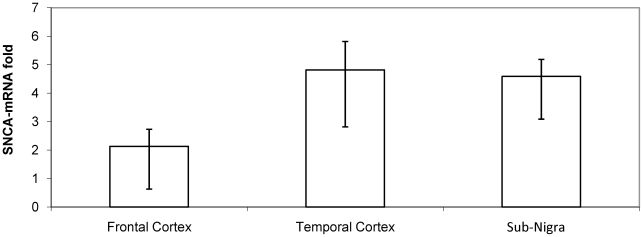
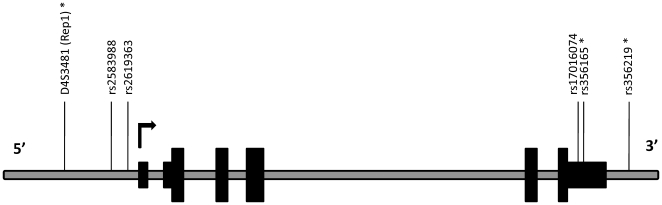
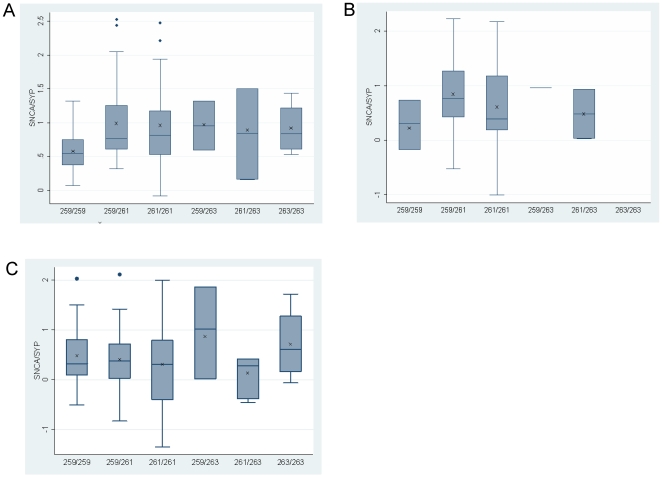
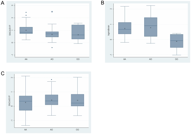
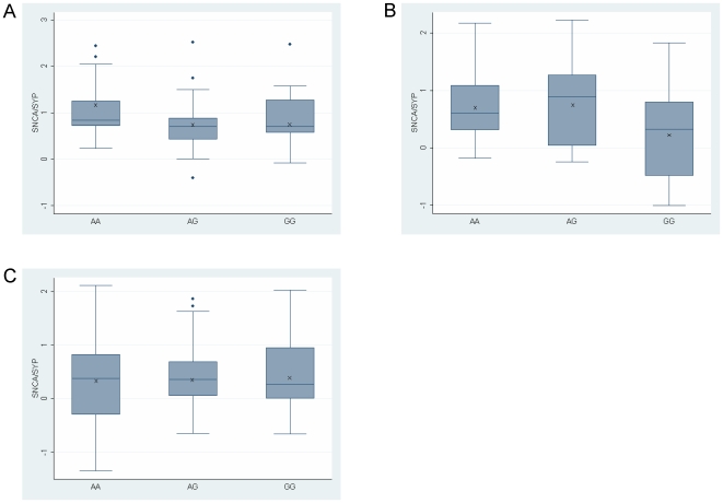
Genetic variability across the SNCA locus has been repeatedly associated with susceptibility to sporadic Parkinson's disease (PD). Accumulated evidence emphasizes the importance of SNCA dosage and expression levels in PD pathogenesis. However whether genetic variability in the SNCA gene modulates the risk to develop sporadic PD via regulation of SNCA expression remained elusive. We studied the effect of PD risk-associated variants at SNCA 5' and 3'regions on SNCA-mRNA levels in vivo in 228 human brain samples from three structures differentially vulnerable to PD pathology (substantia-nigra, temporal- and frontal-cortex) obtained from 144 neurologically normal cadavers. The extensively characterized PD-associated promoter polymorphism, Rep1, had an effect on SNCA-mRNA levels. Homozygous genotype of the 'protective', Rep1-259 bp allele, was associated with lower levels of SNCA-mRNA relative to individuals that carried at least one copy of the PD-risk associated alleles, amounting to an average decrease of approximately 40% and >50% in temporal-cortex and substantia-nigra, respectively. Furthermore, SNPs tagging the SNCA 3'-untranslated-region also showed effects on SNCA-mRNA levels in both the temporal-cortex and the substantia-nigra, although, in contrast to Rep1, the 'decreased-risk' alleles were correlated with increased SNCA-mRNA levels. Similar to Rep1 findings, no difference in SNCA-mRNA level was seen with different SNCA 3'SNP alleles in the frontal-cortex, indicating there is brain-region specificity of the genetic regulation of SNCA expression. We provide evidence for functional consequences of PD-associated SNCA gene variants in disease relevant brain tissues, suggesting that genetic regulation of SNCA expression plays an important role in the development of the disease.


Conflict of interest statement

Competing Interests: The authors have declared that no competing interests exist.

Figures

Figure 1
Figure 1. Average fold expression of SNCA-mRNA in three matched brain regions obtained from the same subjects (n = 7).
Fold levels of SNCA-mRNA were assayed by real-time RT-PCR using TaqMan technology and calculated relative to the geometric mean of SYP- and ENO2- mRNAs reference control using the 2−ΔCt method. The bar graph presents the average of SNCA-mRNA fold expression (mean±S.E.M) of the 7 subjects for each brain region. SN-substantia nigra; TC-temporal cortex; FC- frontal cortex.
Figure 2
Figure 2. A schematic representation of the human SNCA gene with the relative positions of the markers.
Organization of the human SNCA locus: translated exons, wide black solid boxes; 5′ and 3′UTR, narrow black solid boxes; introns and intergenic regions, grey line. The relative positions of the genetic variants are indicated above, asterisks designate variants that were associated with SNCA-mRNA.
Figure 3
Figure 3. Effect of the SNCA-Rep1 promoter genotypes on human SNCA-mRNA expression levels in human brains.
Individuals were genotyped for Rep1. Three brain regions were analyzed: temporal cortex (A), midbrain including SN (B) and frontal cortex (C). In each brain region fold levels of human SNCA-mRNA were assayed by real-time RT-PCR using TaqMan technology and calculated relative to human SYP-mRNA reference control using the 2−ΔCt method. (A) Analysis of the temporal cortex showed that the protective genotype 259/259 correlates with lower SNCA-mRNA levels then the five other genotypes (P = 0.02). The association trend was confirmed in a subset of samples using also the GAPDH and ENO2 reference genes (Table S2). (B) In the midbrain including SN the 259/259 correlates, with lower SNCA-mRNA levels. (C) No correlations of Rep1 genotypes with SNCA-mRNA levels were detected in the frontal cortex. For each genotype the box plot represents the analysis performed using all brain samples available from the specific brain region, each of which was analyzed twice independently, each time in duplicate. The average values are presented in ‘X’. The box plot shows the median (horizontal line inside the box) and the 25th and 75th percentiles (horizontal borders of the box). The range between the 25th and 75th percentiles is the interquartile-range (IQR). The whiskers show the minimal and maximal values inside the main data body.
Figure 4
Figure 4. Effect of SNP rs356219, 3′region genotypes, on human SNCA-mRNA expression levels in human brains.
Individuals were genotyped for SNP rs356219. Three brain regions were analyzed: temporal cortex (A), midbrain including SN (B) and frontal cortex (C). In each brain region fold levels of human SNCA-mRNA were assayed by real-time RT-PCR using TaqMan technology and calculated relative to human SYP-mRNA reference control using the 2-ΔCt method. (A) Analysis of the temporal cortex showed that the protective genotype AA correlates with higher SNCA-mRNA levels than the GA and GG genotypes (P = 0.013). The association trend was confirmed in a subset of samples using also the GAPDH and ENO2 reference genes (Table S2). (B) In the midbrain including SN the AA and AG genotypes correlate with higher SNCA-mRNA levels compared with the GG risk genotype (P<0.05). (C) No correlations of SNP rs356219 genotypes with SNCA-mRNA levels were detected in the frontal cortex. For each genotype the box plot represents the analysis performed using all brain samples available from the specific brain region, each of which was analyzed twice independently, each time in duplicate. The average values are presented in ‘X’. The box plot shows the median (horizontal line inside the box) and the 25th and 75th percentiles (horizontal borders of the box). The range between the 25th and 75th percentiles is the interquartile-range (IQR). The whiskers show the minimal and maximal values inside the main data body.
Figure 5
Figure 5. Effect of SNP rs365165, 3′region genotypes, on human SNCA-mRNA expression levels in human brains.
Individuals were genotyped for SNP rs365165. Three brain regions were analyzed: temporal cortex (A), midbrain including SN (B) and frontal cortex (C). In each brain region fold levels of human SNCA-mRNA were assayed by real-time RT-PCR using TaqMan technology and calculated relative to human SYP-mRNA reference control using the 2-ΔΔCt method. (A) Analysis of the temporal cortex showed that the protective genotype AA correlates with higher SNCA-mRNA levels than the GA and GG genotypes (P<0.05). (B) In the midbrain including SN, the AA and AG genotypes correlate with higher SNCA-mRNA levels compared with the GG risk genotype (P<0.05). (C) No correlations of SNP rs365165 genotypes with SNCA-mRNA levels were detected in the frontal cortex. For each genotype, the box plot represents the analysis performed using all brain samples available from the specific brain region, each of which was analyzed twice independently, each time in duplicate. The average values are presented in ‘X’. The box plot shows the median (horizontal line inside the box) and the 25th and 75th percentiles (horizontal borders of the box). The range between the 25th and 75th percentiles is the interquartile-range (IQR). The whiskers show the minimal and maximal values inside the main data body.

References

    1. Spillantini MG, Schmidt ML, Lee VM, Trojanowski JQ, Jakes R, et al. Alpha-synuclein in Lewy bodies. Nature. 1997;388:839–840. - PubMed
    1. Polymeropoulos MH, Lavedan C, Leroy E, Ide SE, Dehejia A, et al. Mutation in the alpha-synuclein gene identified in families with Parkinson's disease. Science. 1997;276:2045–2047. - PubMed
    1. Kruger R, Kuhn W, Muller T, Woitalla D, Graeber M, et al. Ala30Pro mutation in the gene encoding alpha-synuclein in Parkinson's disease. Nat Genet. 1998;18:106–108. - PubMed
    1. Zarranz JJ, Alegre J, Gomez-Esteban JC, Lezcano E, Ros R, et al. The new mutation, E46K, of alpha-synuclein causes Parkinson and Lewy body dementia. Ann Neurol. 2004;55:164–173. - PubMed
    1. Singleton AB, Farrer M, Johnson J, Singleton A, Hague S, et al. alpha-Synuclein locus triplication causes Parkinson's disease. Science. 2003;302:841. - PubMed

Publication types