

Cytometric assessment of cytostatic and cytotoxic effects of topical glaucoma medications on human epithelial corneal line cells
- PMID: 19834965
- PMCID: PMC3003611
- DOI: 10.1002/cyto.b.20493
Cytometric assessment of cytostatic and cytotoxic effects of topical glaucoma medications on human epithelial corneal line cells
Abstract
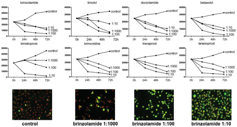
Background: The long-term-treatment of glaucoma with topical medications is associated with side effects involving cornea damage. We examined the effect of glaucoma topical medications (bimatoprost, travoprost, latanoprost, timolol, betaxolol, dorzolamide, brinzolamide, brimonidine) on growth of cells of three human epithelial corneal lines.
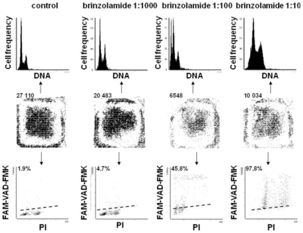
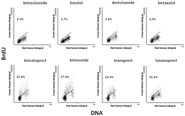
Methods: The cells were cultured in 8-chamber slides, treated with different concentrations of the medications, and fixed at 24, 48, and 72 h. Cell number on slides to estimate viability and growth curves, frequency of apoptosis (FLICA and caspase-3 activation probes), and proliferation (BrdU incorporation assay) were measured by laser scanning cytometry (LSC).
Results: Depending on concentration all examined medications induced cell necrosis or apoptosis and suppressed proliferation. Significant variability in proliferation and apoptosis was observed within the same cultures depending on local cell density, with cells in high density areas being more resistant. The data indicate that commonly used topical medications exert cytostatic and cytotoxic effects in cultures of corneal cells and suggest that caution should be exercised in their use, particularly, when the corneal diseases are accompanied by cell proliferation and regeneration, in long-term-treatment.
Conclusions: The present approach of using LSC makes it possible to assess and compare cytostatic and cytotoxic effects of different topical medications on the respective target cells.
(c) 2009 Clinical Cytometry Society.
Figures

References
-
- Cheong H, Johnson J, Cormier M, Hosseini K. In vitro cytotoxicity of eight β-blockers in human corneal epithelial and retinal pigment epithelial cell lines: Comparison with epidermal keratinocytes and dermal fibroblasts. Toxicol In Vitro. 2008;22:1070–1076. - PubMed
-
- Markstein R. Physiological aspects in the transfer of topical drugs into, within, and out of the eye. In: Orgül S, Flammer J, editors. Pharmacotherapy in Glaucoma. Bern: Verlag Hans Huber; 2000. pp. 55–63.
-
- Okada Y. Effect of topical antiglaucoma medictions on corneal epithelium as evaluation by gene expression patterns. Cornea. 2007;26 (Suppl 1):S46–S54. - PubMed
-
- Noecker RJ, Herrygers LA, Anwaruddin R. Cornea and conjunctival changes caused by commonly used glaucoma medication. Cornea. 2004;23:490–496. - PubMed
-
- Inoue K, Okugawa K, Oshika T, Amano S. Influence of dorzolamide on corneal endotherium. Jpn J Ophthalmol. 2003;47:129–133. - PubMed
Publication types
MeSH terms
Substances
Grants and funding
LinkOut - more resources
Full Text Sources
Medical
Research Materials
