. 2010 Jan;48(1):112-21.
doi: 10.1016/j.yjmcc.2009.09.019. Epub 2009 Oct 14.

A novel computational model of the human ventricular action potential and Ca transient

Affiliations

A novel computational model of the human ventricular action potential and Ca transient

Eleonora Grandi et al. J Mol Cell Cardiol. 2010 Jan.

Abstract

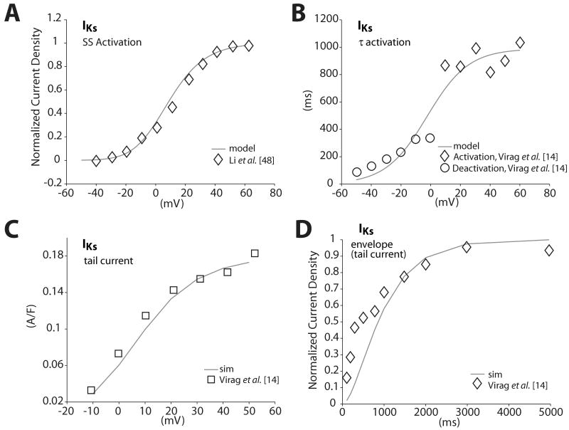
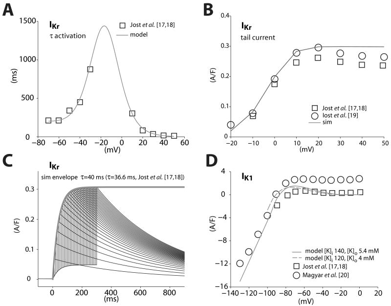
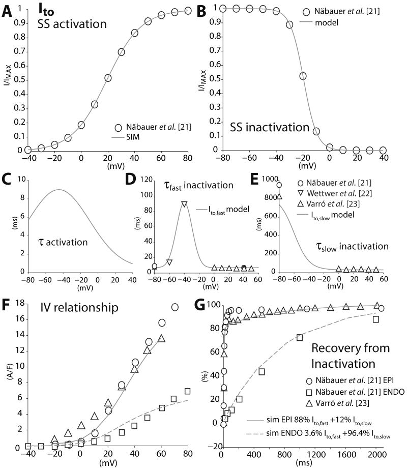
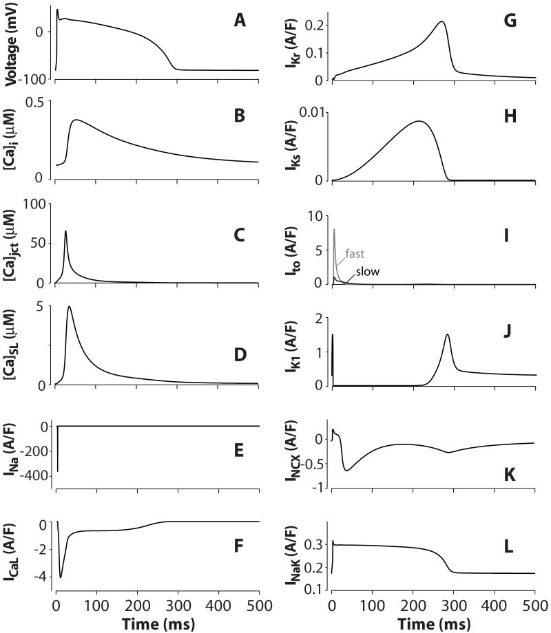
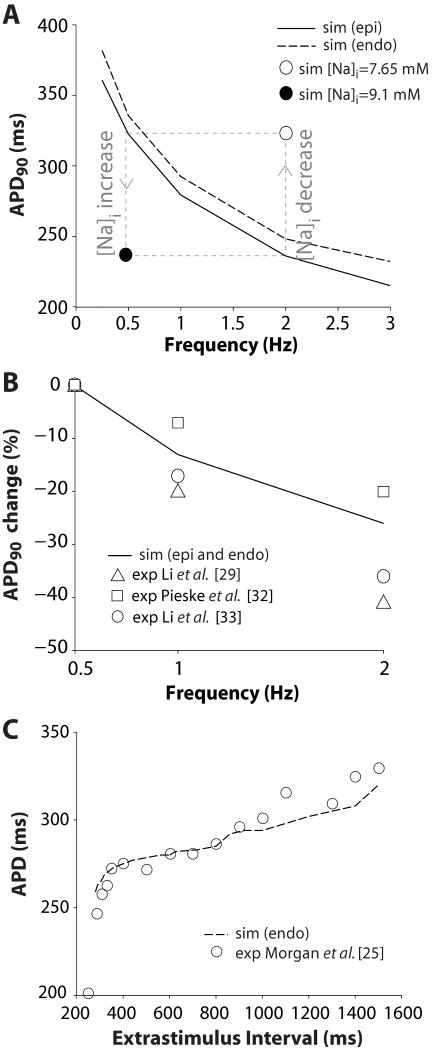
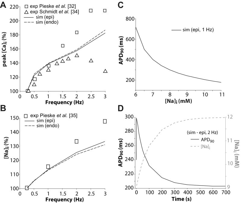
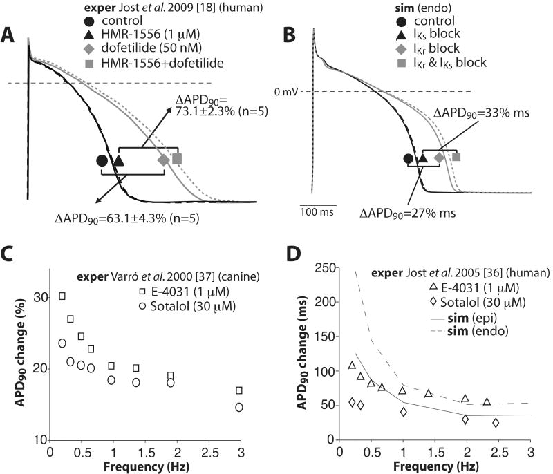
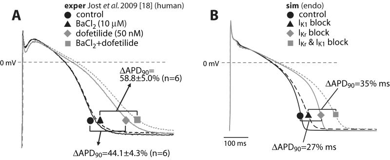
We have developed a detailed mathematical model for Ca handling and ionic currents in the human ventricular myocyte. Our aims were to: (1) simulate basic excitation-contraction coupling phenomena; (2) use realistic repolarizing K current densities; (3) reach steady-state. The model relies on the framework of the rabbit myocyte model previously developed by our group, with subsarcolemmal and junctional compartments where ion channels sense higher [Ca] vs. bulk cytosol. Ion channels and transporters have been modeled on the basis of the most recent experimental data from human ventricular myocytes. Rapidly and slowly inactivating components of I(to) have been formulated to differentiate between endocardial and epicardial myocytes. Transmural gradients of Ca handling proteins and Na pump were also simulated. The model has been validated against a wide set of experimental data including action potential duration (APD) adaptation and restitution, frequency-dependent increase in Ca transient peak and [Na](i). Interestingly, Na accumulation at fast heart rate is a major determinant of APD shortening, via outward shifts in Na pump and Na-Ca exchange currents. We investigated the effects of blocking K currents on APD and repolarization reserve: I(Ks) block does not affect the former and slightly reduces the latter; I(K1) blockade modestly increases APD and more strongly reduces repolarization reserve; I(Kr) blockers significantly prolong APD, an effect exacerbated as pacing frequency is decreased, in good agreement with experimental results in human myocytes. We conclude that this model provides a useful framework to explore excitation-contraction coupling mechanisms and repolarization abnormalities at the single myocyte level.


Figures

Figure 1
Figure 1
Gating properties of IKs. Model curves (lines) and data (symbols) from Li et al. [48] and Virág et al. [14] are shown. A: Steady-state activation. B: Activation and deactivation time constants. C: Current-voltage relationship. Tail IKs was measured during 5,000-ms voltage steps to potentials shown from a holding potential of −40 mV. D: Envelope test protocol assessing the kinetics of IKs activation.
Figure 2
Figure 2
Gating properties of IKr and IK1. Model curves (lines) and data (symbols) from Jost et al. [17, 18], Iost et al. [19] and Magyar et al. [20] are shown. A: Activation and deactivation time constants. B: IKr current-voltage relationship. Tail IKr was measured during 1,000-ms voltage steps to potentials shown from a holding potential of −40 mV. C: Simulated envelope test protocol assessing IKr activation time constant at +30 mV. D: Steady-state current-voltage relationships of IK1.
Figure 3
Figure 3
Gating properties of Ito. Model curves (lines) and data (symbols) from Näbauer et al. [21], Wettwer et al. [22] and Varró et al. [23] are shown. A: Steady-state activation. B: Steady-state inactivation. C: Activation time constants of Ito,fast and Ito,slow are unchanged from Shannon et al. [2]. D: Inactivation time constants of Ito,fast and E: Ito,slow. F: Epicardial and endocardial Ito current-voltage curves. G: Recovery from inactivation of epicardial and endocardial Ito was assessed with a two pulse (500 ms from −80 to 50 mV) protocol with varying interpulse intervals.
Figure 4
Figure 4
Steady-state epicardial AP, calcium transient, and major ionic currents simulated at 1-Hz pacing rate. A: Action potential. B: Cytosolic [Ca]. C: Junctional [Ca]. D: SL [Ca]. E: Fast sodium current. F: L-type calcium current. G: Rapidly activating delayed rectifier potassium current. H: Slowly activating delayed rectifier potassium current. I: Slowly and rapidly inactivating transient outward potassium currents. J: Inward rectifier potassium current. K: Sodium-calcium exchange current. L: Sodium-potassium ATPase current.
Figure 5
Figure 5
A: Simulated APD adaptation curves. Steady-state APD of human epicardial (solid line) and endocardial (dashed line) ventricular cell models shortened as pacing rate was made faster. B: Fractional APD90 changes on increasing pacing frequency from 0.5 to 1 and 2 Hz. Simulated results (solid line) are within the range of experimental variability (data are from references [29, 32, 33], symbols). C: Simulated endocardial APD restitution curve (dashed line), compared with experimental data (circles) from endocardial monophasic APs in human hearts [25].
Figure 6
Figure 6
A: Fractional changes in peak [Ca]i and B: [Na]i at increasing pacing frequencies in the human epicardial (solid lines) and endocardial (dashed lines) ventricular myocyte models. Experimental results (symbols) are from references [32, 34] and [35]. C: Predicted APD90 dependency on [Na]i (epicardium, 1 Hz). D: Simulated Na pump blockade (50%) caused AP prolongation followed by gradual shortening as Na ions accumulated in the cytosol (epicardium, 2 Hz).
Figure 7
Figure 7
Contribution of IKs and IKr to ventricular repolarization and repolarization reserve. A: Effects of IKs (HMR-1556), IKr (dofetilide), and combined (IKs+IKr) block on APD in isolated human myocytes. Redrawn from [18]. B: Simulations predicted a large effect of IKr block on APD, no effect of IKs on basal APD and modest contribution to repolarization reserve. C, D: IKr blockers prolonged APD in a reverse-frequency-dependent manner. Model predictions (D, lines) agree with experimental findings (symbols) in (C) canine [37] and (D) human ventricular myocytes [36].
Figure 8
Figure 8
Contribution of IK1 to ventricular repolarization and repolarization reserve. A: Effect of IK1 (BaCl2), IKr (dofetilide) and combined (IK1+IKr) block on APD in isolated human myocytes. Redrawn from [18]. B: Simulated results predict a modest AP prolongation upon IK1 block and a more significant contribution of IK1 to repolarization reserve.

References

    1. Faber GM, Rudy Y. Action Potential and Contractility Changes in [Na+]i Overloaded Cardiac Myocytes: A Simulation Study. Biophys J. 2000;78(5):2392. - PMC - PubMed
    1. Shannon TR, Wang F, Puglisi J, Weber C, Bers DM. A Mathematical Treatment of Integrated Ca Dynamics within the Ventricular Myocyte. Biophys J. 2004;87(5):3351. - PMC - PubMed
    1. Bondarenko VE, Szigeti GP, Bett GCL, Kim SJ, Rasmusson RL. Computer model of action potential of mouse ventricular myocytes. Am J Physiol Heart Circ Physiol. 2004;287(3):H1378–1403. - PubMed
    1. Hund TJ, Rudy Y. Rate Dependence and Regulation of Action Potential and Calcium Transient in a Canine Cardiac Ventricular Cell Model. Circulation. 2004;110(20):3168–3174. - PMC - PubMed
    1. Grandi E, Puglisi JL, Wagner S, Maier LS, Severi S, Bers DM. Simulation of Ca-Calmodulin-Dependent Protein Kinase II on Rabbit Ventricular Myocyte Ion Currents and Action Potentials. Biophys J. 2007;93(11):3835. - PMC - PubMed

Publication types