. 2009 Oct 30;139(3):485-98.
doi: 10.1016/j.cell.2009.09.033.

Induction of intestinal Th17 cells by segmented filamentous bacteria

Affiliations

Induction of intestinal Th17 cells by segmented filamentous bacteria

Ivaylo I Ivanov et al. Cell. .

Abstract

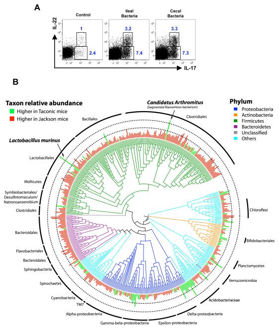
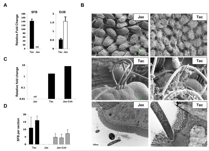
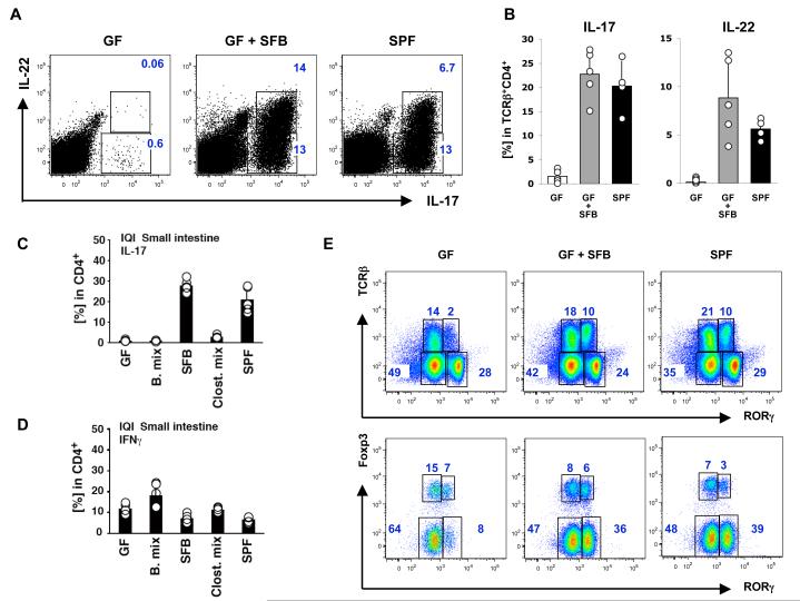
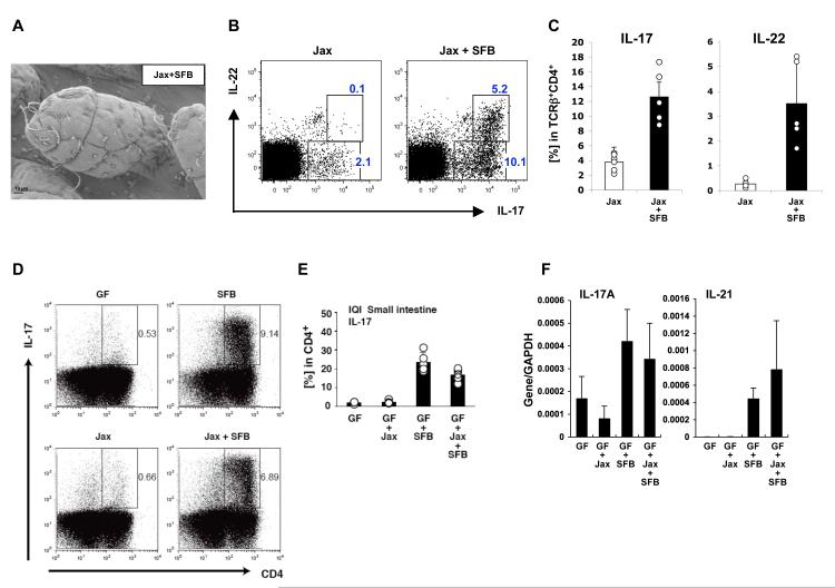
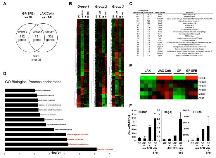
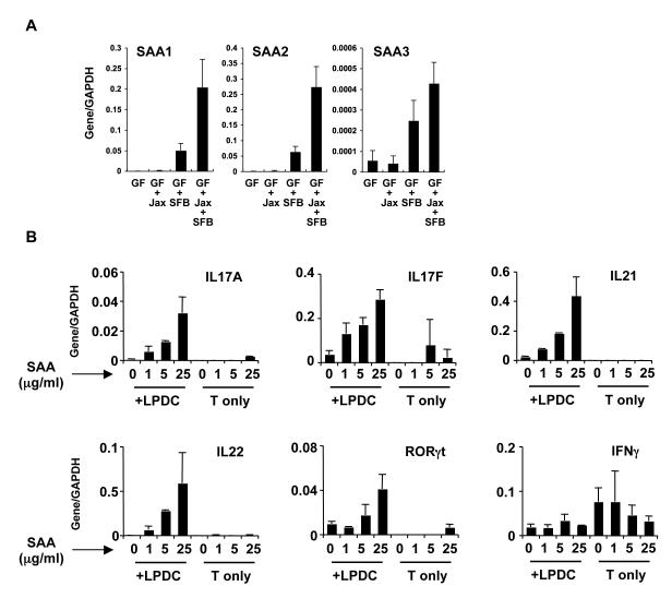
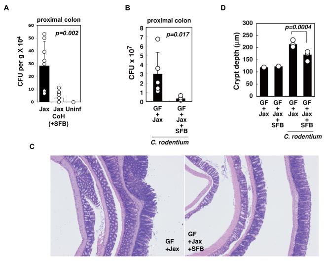
The gastrointestinal tract of mammals is inhabited by hundreds of distinct species of commensal microorganisms that exist in a mutualistic relationship with the host. How commensal microbiota influence the host immune system is poorly understood. We show here that colonization of the small intestine of mice with a single commensal microbe, segmented filamentous bacterium (SFB), is sufficient to induce the appearance of CD4(+) T helper cells that produce IL-17 and IL-22 (Th17 cells) in the lamina propria. SFB adhere tightly to the surface of epithelial cells in the terminal ileum of mice with Th17 cells but are absent from mice that have few Th17 cells. Colonization with SFB was correlated with increased expression of genes associated with inflammation and antimicrobial defenses and resulted in enhanced resistance to the intestinal pathogen Citrobacter rodentium. Thus, manipulation of this commensal-regulated pathway may provide new opportunities for enhancing mucosal immunity and treating autoimmune disease.


Figures

Figure 1
Figure 1. Comparative analysis of the microbiota in the terminal ileum of C57BL/6 (B6) mice from Jackson Laboratory versus Taconic Farms
A. Lumenal bacteria from both cecum and terminal ileum can induce Th17 cell differentiation upon transfer into Jackson B6 mice. Jackson B6 mice were gavaged with water (control) or with intestinal luminal contents from cecum or terminal ileum of Taconic B6 mice. LPL from small intestine were isolated 10 days later and analyzed for intracellular cytokines. Representative plots from one experiment with 3 mice per group. Plots gated on TCRβ+CD4+ LPL. B. Phylogenetic tree based on 16S rRNA gene sequences of bacterial taxa detected in the terminal ileum showing significantly different relative abundances (PhyloChip fluorescence intensity) between the suppliers, Taconic and Jackson. Branches of the tree are color coded according to phylum while green and red bars display taxa with significantly greater relative abundance in Taconic and Jackson mice, respectively. The inner and outer dotted rings represents intensities corresponding to 5-fold and 25-fold differences in 16S copy number. The two taxa with the greatest difference between Taconic and Jackson mice, Lactobacillus murinus (~94-fold difference) and Candidatus Arthromitus (~40-fold difference), are noted by arrows.
Figure 2
Figure 2. Segmented Filamentous Bacteria (SFB) in the intestinal tract of Th17 cell-sufficient and Th17 cell-deficient mice
A. Quantitative PCR (qPCR) analysis of SFB and total bacterial (EUB) 16S rRNA genes in mouse feces from Taconic (Tac) and Jackson (Jax) B6 mice. Genomic DNA was isolated from combined fecal pellets from 4 animals from each strain. The experiment was repeated numerous times with similar results. B. Scanning (SEM) and transmission (TEM) electron microscopy of terminal ileum of 8 week-old Jackson (Jax) and Taconic (Tac) C57BL/6 mice housed under similar conditions and diet for at least one week. Note the presence of long filamentous bacteria with SFB morphology in Taconic, but not Jackson mice C. qPCR analysis for SFB presence in Jackson B6 mice after 14 days of co-housing with Taconic B6 mice (Jax-Coh). Genomic DNA was isolated from pooled feces from 3-4 mice per group. D. SFB colonization of terminal ileum of Jackson B6 mice after 14 days of co-housing with Taconic B6 mice (Jax-Coh). Toluidine-blue sections were prepared from 0.5 cm piece of the terminal ileum as described in Methods and examined by light microscopy. Adherent bacteria with SFB morphology were counted in 4-5 sections from each sample. Each column represents a separate animal.
Figure 3
Figure 3. SFB specifically induce Th17 cell differentiation in germ-free mice
A-B. 6-week old Swiss-Webster (SW) germ-free mice (GF) were colonized with SFB (GF+SFB) as described in Methods and small intestinal lamina propria lymphocytes (SI LPL) were isolated 10 days later. Representative plots in (A) and combined data in (B) of IL-17 and IL-22 expression in TCRβ+CD4+ LPL. Data are from one of three separate experiments with similar results. Error bars (SD). SPF – mice raised under conventional specific pathogen free conditions. Each circle in (B) represents a separate animal C-D. IL-17 (C) and IFNγ (D) expression in TCRβ+CD4+ SI LPL from mice colonized with different commensal bacteria. IQI germ-free (GF) mice were colonized with 16 strains of Bacteroidaceae (B. mix), SFB, 46 strains of Clostridium sp mixture (Clost. mix), or microbiota from conventionally raised mice (SPF). Intracellular cytokine production in SI LP CD4 T cells was analyzed 3 weeks later by flow cytometry. Circles represent separate animals E. SFB colonization induces RORγt expression only in CD4+ T cells. RORγt expression in total SI LPL (top panels) and RORγt and Foxp3 expression in TCRβ+CD4+ SI LPL (bottom panels) in GF mice, GF mice colonized with SFB (GF+SFB) and conventionally raised mice (SPF).
Figure 4
Figure 4. SFB induce Th17 cell differentiation upon colonization of Jackson C57BL/6 mice
A. Colonization of the terminal ileum by SFB 10 days after transfer of fecal homogenates from SFB-mono mice into Jackson B6 mice B-C. IL-17 and IL-22 expression in TCRβ+CD4+ SI LPL in Jackson B6 mice colonized with SFB (Jax+SFB) compared to controls (Jax). Data from one of two experiments D-E. Jackson microbiota induces Th17 cells only when complemented with SFB. Germ-free IQI mice were colonized with SFB, Jackson microbiota isolated from fecal pellets by itself (Jax), or a mixture of both (Jax + SFB). Th17 cell proportions were analyzed in the LP 3 weeks later by flow cytometry. Plots in (D) gated on total lymphocytes. Data in (E) represent percentage of IL-17+ cells in the CD4+ gate F. RT-PCR for Th17 cell effector cytokines in total RNA from terminal ileum of the mice in (E)
Figure 5
Figure 5. Transcriptional programs induced by SFB colonization
A. Venn diagrams showing the overlap between genes affected by either SFB colonization of SW GF mice only (Group 2), introduction of Taconic microbiota into Jackson B6 mice by co-housing (Group 1), or both (Group 3). Total RNA was prepared from terminal ileum of the corresponding mice after 10 days of colonization and Affymetrix gene chip analysis was performed as described in Methods. B. Heat-map analysis of the three groups in (A). Each line represents a single Affymetrix probe and each column a single mouse. Green, probes that were at least 2 fold down-regulated. Red, probes that were at least 2 fold up-regulated. C. Top up-regulated genes in Group 3 in (A) arranged by fold change in GF+SFB mice. D. Biological processes specifically induced by SFB (genes in Group 3 in (A)). Gene ontology analysis was performed as described in Methods. E. Changes in anti-microbial peptide related genes upon SFB colonization and Th17 cell induction by co-housing. Each column represents an individual mouse. F. RT-PCR analysis of selected genes, induced by SFB colonization. IQI GF mice (GF) were colonized with fecal homogenates from SFB-mono mice (SFB), Jackson B6 mice (Jackson), or a mixture of both (Jackson+SFB). Total RNA from terminal ileum was prepared 3 weeks later and RT-PCR performed as described in Methods.
Figure 6
Figure 6. SFB colonization induces SAA expression that influences Th17 differentiation
A. Relative mRNA expression levels of SAA1-3 genes by real-time RT-PCR in the terminal ileum of IQI GF mice (GF) colonized with fecal homogenates from SFB-mono mice (SFB), Jackson B6 mice (Jackson), or a mixture of both (Jackson+SFB). B. Splenic naïve CD4+ T cells were cocultured with or without LP CD11c+ cells in the presence of an anti-CD3 antibody with the indicated concentration of recombinant Apo-SAA for 4 days. T cells were collected, restimulated with PMA and ionomycin, and real-time RT-PCR performed. Results were normalized to expression of GAPDH mRNA. The data are representative of four independent experiments with similar results.
Figure 7
Figure 7. SFB colonization confers protection from infection by Citrobacter rodentium
A. Jackson B6 mice (Jax) were co-housed with Taconic B6 mice (Jax CoH) for 10 days to induce colonization with SFB and Th17 cells. Both groups were infected with ~ 1 × 10⁹ CFU of C. rodentium/mouse and pathogen colonization of the colon was examined at day 8 of infection (n = 9/group). Uninf – uninfected controls. B-D. IQI GF mice colonized with Jackson microbiota (Jax) or Jax+SFB were orally infected with 2 × 10⁹ CFU of C. rodentium, and colons were harvested at day 8 of infection (n= 5/group). C. rodentium CFUs in proximal colon (B), histopathology (C) and crypt length (D) in the distal colon (H&E). Data represent means ± SD and circles represent separate animals. Similar effects of SFB on C. rodentium colonization were observed in a separate experiment with C.B17 mice colonized with ASF with and without SFB.

Comment in

References

    1. Abbas AK, Murphy KM, Sher A. Functional diversity of helper T lymphocytes. Nature. 1996;383:787–793. - PubMed
    1. Atarashi K, Nishimura J, Shima T, Umesaki Y, Yamamoto M, Onoue M, Yagita H, Ishii N, Evans R, Honda K, et al. ATP drives lamina propria T(H)17 cell differentiation. Nature. 2008;455:808–812. - PubMed
    1. Aujla SJ, Dubin PJ, Kolls JK. Th17 cells and mucosal host defense. Semin Immunol. 2007;19:377–382. - PMC - PubMed
    1. Backhed F, Ley RE, Sonnenburg JL, Peterson DA, Gordon JI. Host-bacterial mutualism in the human intestine. Science. 2005;307:1915–1920. - PubMed
    1. Barman M, Unold D, Shifley K, Amir E, Hung K, Bos N, Salzman N. Enteric salmonellosis disrupts the microbial ecology of the murine gastrointestinal tract. Infect Immun. 2008;76:907–915. - PMC - PubMed

Publication types

MeSH terms

Associated data