Review
. 2009 Nov 20;31(5):711-21.
doi: 10.1016/j.immuni.2009.09.010.

Chemokines and chemokine receptors: standing at the crossroads of immunobiology and neurobiology

Affiliations
Review

Chemokines and chemokine receptors: standing at the crossroads of immunobiology and neurobiology

Richard M Ransohoff. Immunity. .

Abstract

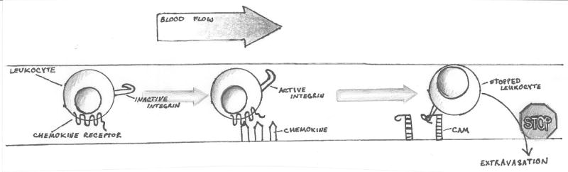
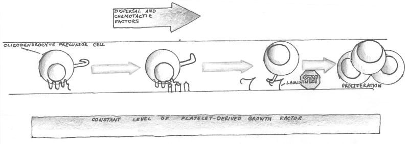
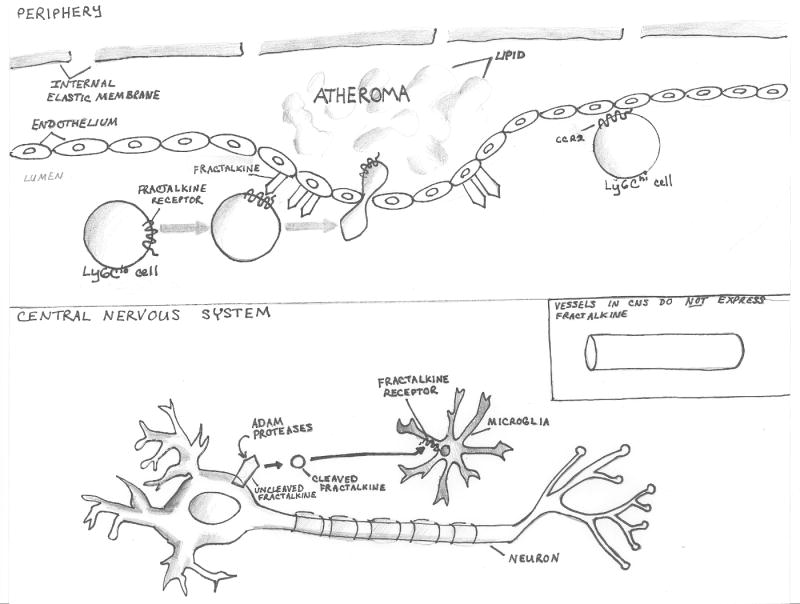
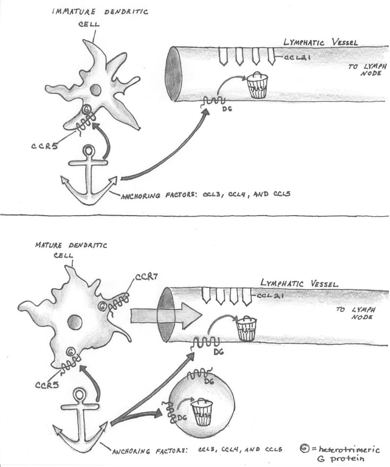
There are several molecular entities common to the immune and nervous systems. Salient among them are the chemokines and their receptors, which play remarkably varied and potent roles in immunobiology and neurobiology. This review limns several illustrative examples and presents general principles of chemokine action that are manifest in both systems. These include the following: (1) chemokines tend equally to arrest cells and to make them move, in the process of positioning target cells with spatiotemporal precision; (2) signaling and nonsignaling receptors collaborate to adjust the chemokine environment for maximal efficacy; and (3) expression of a single chemokine receptor on circulating blood cells and parenchymal cells is often used to coordinate complex tissue responses. The challenge is to integrate knowledge of the roles of key receptors (as well as their ligands) into a coherent account of events during pathologic processes, in order to guide therapeutic development.


Figures

Figure 1
Figure 1
Figure 1a. Chemokine receptor signaling mediates arrest of moving cells in the immune system. The cartoon illustrates the function of chemokine receptor signaling in the early phases of leukocyte extravasation under flow. Initial leukocyte-endothelial interactions are loose and reversible. Chemokines are immobilized in the vascular lumen by interactions with glycosaminoglycans or by presentation through molecules such as the Duffy Antigen Receptor for Chemokines (DARC). Chemokine receptor signaling converts inactive integrin to active integrin, capable of high-affinity interaction with Cell Adhesion Molecules (CAMs) on the endothelial luminal surface. This interaction arrests the leukocyte under flow and extravasation of the leukocyte often follows. Figure 1b. Chemokine receptor signaling mediates arrest of moving cells in the developing nervous system. The lower panel shows an oligodendrocyte precursor cell (OPC) moving through the presumptive white matter of the post-natal rodent spinal cord. Chemokine CXCL1 is expressed focally in these tissues and signals to CXCR2 on the OPC. In an in-vitro model, signaling through CXCR2 leads to increased interaction between the OPC and a laminin substrate. Studies using purified OPCs in vitro, and in vivo analysis of CXCR2-/- mice suggest that arrest of the OPC in a high local concentration of CXCL1 and PDGF (which is present throughout the developing spinal cord) drives a burst of OPC proliferation.
Figure 1
Figure 1
Figure 1a. Chemokine receptor signaling mediates arrest of moving cells in the immune system. The cartoon illustrates the function of chemokine receptor signaling in the early phases of leukocyte extravasation under flow. Initial leukocyte-endothelial interactions are loose and reversible. Chemokines are immobilized in the vascular lumen by interactions with glycosaminoglycans or by presentation through molecules such as the Duffy Antigen Receptor for Chemokines (DARC). Chemokine receptor signaling converts inactive integrin to active integrin, capable of high-affinity interaction with Cell Adhesion Molecules (CAMs) on the endothelial luminal surface. This interaction arrests the leukocyte under flow and extravasation of the leukocyte often follows. Figure 1b. Chemokine receptor signaling mediates arrest of moving cells in the developing nervous system. The lower panel shows an oligodendrocyte precursor cell (OPC) moving through the presumptive white matter of the post-natal rodent spinal cord. Chemokine CXCL1 is expressed focally in these tissues and signals to CXCR2 on the OPC. In an in-vitro model, signaling through CXCR2 leads to increased interaction between the OPC and a laminin substrate. Studies using purified OPCs in vitro, and in vivo analysis of CXCR2-/- mice suggest that arrest of the OPC in a high local concentration of CXCL1 and PDGF (which is present throughout the developing spinal cord) drives a burst of OPC proliferation.
Figure 2
Figure 2. Differential expression of fractalkine/CX3CL1 in the peripheral vascular system and in the central nervous system (CNS) leads to disparate functional roles
In the periphery (upper panel), fractalkine is expressed on the endothelium as a single-pass transmembrane component, which can mediate firm arrest under flow. Ly6Clo monocytes express high levels of CX3CR1/fractalkine receptor, and can be recruited into a developing atheroma through CX3CL1/CX3CR1 interactions. Both CX3CR1-/- mice and individuals harboring a polymorphic variant of CX3CR1 (CX3CR1I249/M280) which acts as a dominant negative inhibitor of signaling are relatively protected from atherosclerosis. Atheromata are also infiltrated by Ly6Chi monocytes via signaling to CCR2. Fractalkine is not present on CNS vessels. Instead, fractalkine is expressed by neurons, where it can be cleaved and released by ADAM proteases, so that the healthy adult mouse brain contains 150-300pg/mg protein of soluble fractalkine. Microglia uniformly express CX3CR1 and respond to ligand by modulating effector functions in a manner that limits toxicity to neurons. Individuals bearing the CX3CR1I249/M280 variant are at increased risk for Age-related Macular Degeneration (AMD), an inflammatory neurodegenerative disorder, possibly because of neurotoxicity mediated by retinal microglia.
Figure 3
Figure 3
Figure 3A-B. Nonsignaling ‘scavenger’ chemokine receptor-like molecules adjust the chemokine environment to optimize immune function. In inflamed tissues, an immature dendritic cell (3A) encounters a high concentration of chemokines (CCL3, CCL4, CCL5) which all signal to CCR5 and ‘anchor’ the cell in place, while it's processing antigen and upregulating functions associated with antigen presentation. During this time, D6, a nonsignaling scavenger receptor on lymphatic endothelium, clears excess inflammatory CC chemokines. After maturation (3B), the dendritic cell downregulates CCR5, limiting signaling from CCL3, CCL4 and CCL5. The mature dendritic cell also upregulates CCR7. At the same time, other leukocytes upregulate D6 and assist in removing these inflammatory chemokines (but not the homeostatic chemokine CCL21) from the environment. The culmination of reduced signaling from inflammatory chemokines and suppressed signaling from inflammatory chemokines favors entry of the dendritic cell into the afferent lymphatic vessel, whose lumen is decorated with CCL21. The dendritic cell will subsequently traffick to the draining lymph node. Figure 3C. Nonsignaling ‘scavenger’ chemokine receptor-like molecules adjust the chemokine environment to optimize neuron precursor migration during development. The lateral line primordium comprises a population of cells, organized in a coherent linear aggregate, whose migration is mediated by chemokine CXCL12. Cells at the leading edge express the signaling receptor CXCR4. Cells at the trailing edge express CXCR7 and enhance gradient steepness, by removing CXCL12.
Figure 3
Figure 3
Figure 3A-B. Nonsignaling ‘scavenger’ chemokine receptor-like molecules adjust the chemokine environment to optimize immune function. In inflamed tissues, an immature dendritic cell (3A) encounters a high concentration of chemokines (CCL3, CCL4, CCL5) which all signal to CCR5 and ‘anchor’ the cell in place, while it's processing antigen and upregulating functions associated with antigen presentation. During this time, D6, a nonsignaling scavenger receptor on lymphatic endothelium, clears excess inflammatory CC chemokines. After maturation (3B), the dendritic cell downregulates CCR5, limiting signaling from CCL3, CCL4 and CCL5. The mature dendritic cell also upregulates CCR7. At the same time, other leukocytes upregulate D6 and assist in removing these inflammatory chemokines (but not the homeostatic chemokine CCL21) from the environment. The culmination of reduced signaling from inflammatory chemokines and suppressed signaling from inflammatory chemokines favors entry of the dendritic cell into the afferent lymphatic vessel, whose lumen is decorated with CCL21. The dendritic cell will subsequently traffick to the draining lymph node. Figure 3C. Nonsignaling ‘scavenger’ chemokine receptor-like molecules adjust the chemokine environment to optimize neuron precursor migration during development. The lateral line primordium comprises a population of cells, organized in a coherent linear aggregate, whose migration is mediated by chemokine CXCL12. Cells at the leading edge express the signaling receptor CXCR4. Cells at the trailing edge express CXCR7 and enhance gradient steepness, by removing CXCL12.

Similar articles

Cited by

References

    1. Abbadie C. Chemokines, chemokine receptors and pain. Trends Immunol. 2005;26:529–534. - PubMed
    1. Abbadie C, Lindia JA, Cumiskey AM, Peterson LB, Mudgett JS, Bayne EK, DeMartino JA, Macintyre DE, Forrest MJ. Impaired neuropathic pain responses in mice lacking the chemokine receptor CCR2. Proc Natl Acad Sci U S A. 2003;100:7947–7952. - PMC - PubMed
    1. Acosta JC, O'Loghlen A, Banito A, Guijarro MV, Augert A, Raguz S, Fumagalli M, Da CM, Brown C, Popov N, Takatsu Y, Melamed J, dda di FF, Bernard D, Hernando E, Gil J. Chemokine signaling via the CXCR2 receptor reinforces senescence. Cell. 2008;133:1006–1018. - PubMed
    1. Allen NJ, Attwell D. A chemokine-glutamate connection. Nat Neurosci. 2001;4:676–678. - PubMed
    1. Alon R, Feigelson S. From rolling to arrest on blood vessels: leukocyte tap dancing on endothelial integrin ligands and chemokines at sub-second contacts. Semin Immunol. 2002;14:93–104. - PubMed

Publication types