. 2009 Dec 3;8(12):1400-10.
doi: 10.1016/j.dnarep.2009.09.009.

Adenine removal activity and bacterial complementation with the human MutY homologue (MUTYH) and Y165C, G382D, P391L and Q324R variants associated with colorectal cancer

Affiliations

Adenine removal activity and bacterial complementation with the human MutY homologue (MUTYH) and Y165C, G382D, P391L and Q324R variants associated with colorectal cancer

Sucharita Kundu et al. DNA Repair (Amst). .

Abstract

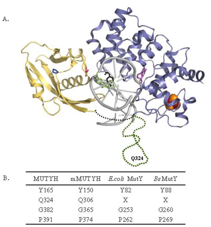
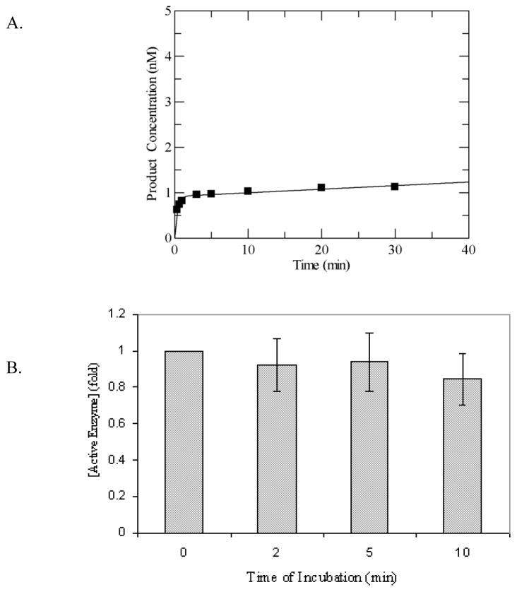
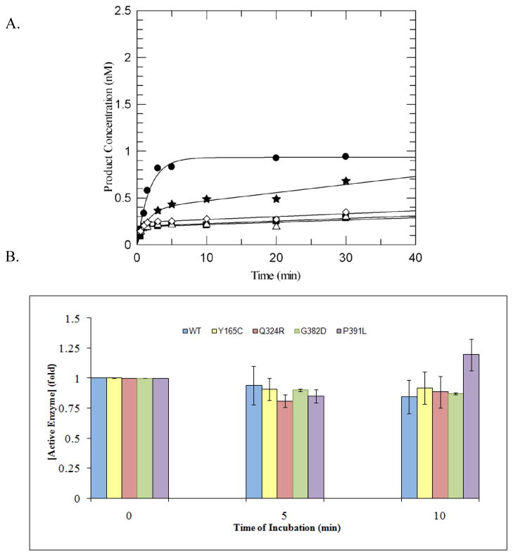
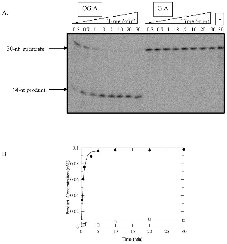
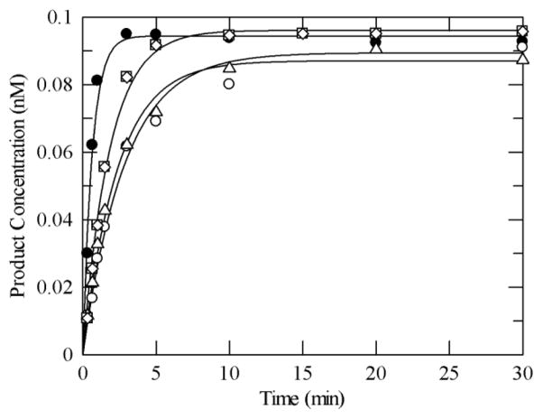
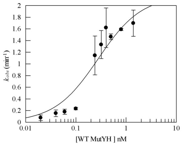
MUTYH-associated polyposis (MAP) is the only inherited colorectal cancer syndrome that is associated with inherited biallelic mutations in a base excision repair gene. The MUTYH glycosylase plays an important role in preventing mutations associated with 8-oxoguanine (OG) by removing adenine residues that have been misincorporated opposite OG. MAP-associated mutations are present throughout MUTYH, with a large number coding for missense variations. To date the available information on the functional properties of MUTYH variants is conflicting. In this study, a kinetic analysis of the adenine glycosylase activity of MUTYH and several variants was undertaken using a correction for active fraction to control for differences due to overexpression and purification. Using these methods, the rate constants for steps involved in the adenine removal process were determined for the MAP variants Y165C, G382D, P391L and Q324R MUTYH. Under single-turnover conditions, the rate of adenine removal for these four variants was found to be 30-40% of WT MUTYH. In addition, the ability of MUTYH and the variants to suppress mutations and complement for the absence of MutY in Escherichia coli was assessed using rifampicin resistance assays. The presence of WT and Q324R MUTYH resulted in complete suppression of the mutation frequency, while G382D MUTYH showed reduced ability to suppress the mutation frequency. In contrast, the mutation frequency observed upon expression of P391L and Y165C MUTYH were similar to the controls, suggesting no activity toward preventing DNA mutations. Notably, though all variations studied herein resulted in similar reductions in adenine glycosylase activity, the effects in the bacterial complementation are quite different. This suggests that the consequences of a specific amino acid variation on overall repair in a cellular context may be magnified.


Figures

Figure 1
Figure 1
MUTYH variations associated with MAP. (A) Bacillus stearothermophilus MutY-DNA cocrystal structure (1RRQ) with residues corresponding to variations in MUTYH involved in MAP highlighted: Y88, black; G260, pink; P269, dark blue; FeS cluster; brown and orange; adenine, violet; OG, green; DNA, grey; N-terminal domain, light blue; C-terminal domain, yellow, linker region between domains, black dotted line. The sequence present only in MUTYH shown in green dotted line harbors position of Q324. This region is not based on the structure, and was added into a structure generated from coordinates (1RRQ) from the reported structures. (B) Summary of conserved residues across different species. X denotes no matching residue found on sequence alignment using Clustal W.
Figure 2
Figure 2
Adenine glycosylase assays of WT MUTYH under multiple-turnover conditions (A) Representative plot of adenine removal activity by WT MUTYH under multiple-turnover conditions at 37 °C and 150 mM buffer salt concentration with an OG:A-containing duplex DNA substrate (10 nM). Line represents fit to equation 1 to determine A0 and k3, which are = 0.92 and 0.008 min−1, respectively, for this particular data set. (B) Stability assays of WT MUTYH based on A0 values determined after incubation of WT MUTYH in assay buffer show no significant reduction in enzyme activity as a function of time. The activity of MUTYH without prior incubation was normalized to 1. Aliquots were removed at intervals of 2, 5 and 10 minutes for adenine glycosylase assays under multiple-turnover conditions, and active enzyme concentrations were determined from the burst amplitudes of the progress curves. The adenine glycosylase activity at each time point was measured in at least three separate experiments. The grey bars represent the average on the basis of the initial amplitude and the error bars represent the standard deviation from the average (as a percentage of the normalized value).
Figure 3
Figure 3
Comparison of burst amplitudes of MUTYH variants to the WT enzyme. (A) Representative plot of adenine removal activity by WT MUTYH and variants under multiple-turnover conditions at 37 °C and 150 mM buffer salt concentration with an OG:A containing duplex DNA substrate (10 nM). WT MUTYH (closed circles), Y165C (closed squares), Q324R (closed stars), G382D (open triangles), P391L (open rhombus) were added to the reaction mix to provide equal total protein concentrations (30 ug as measured by Bradford assay). Lines represent fit to equation 1, with values of A0 obtained for the various enzymes of WT = 0.93; Y165C = 0.19; Q324R= 0.38; G382D = 0.19; P391L = 0.24. (B) Stability assays of WT MUTYH and variants in assay buffer as measured by glycosylase assays show no significant reduction in enzyme activity as a function of time. Aliquots were removed at intervals of 0, 5 and 10 minutes for adenine glycosylase assays under multiple-turnover conditions and fit to equation 1 to determine the burst amplitudes, A0. The activity of MUTYH without prior incubation was normalized to 1. The bars at 5 and 10 min represent the average loss of activity on incubation. The adenine glycosylase activity at each time point was measured in at least three separate experiments and the error bars represent one standard deviation from the average (as a percentage of the normalized value).
Figure 4
Figure 4
Adenine glycosylase assays of MUTYH under single-turnover conditions with OG:A and G:A substrates. (A) Representative storage phosphor autoradiogram of denaturing PAGE experiment. Bands derived from substrate and product are shown. The minus (−) lane represents the G:A-containing DNA with no enzyme added as a control. (B) Plot of adenine removal activity by WT MUTYH with OG:A- (close circles) and G:A- (open squares) containing substrates at 37 °C. Lines represent fits of the data to a single exponential (equation 2). For this particular experiment, k2 = 1.5 min−1 and <0.002 min−1, for OG:A and G:A, respectively. Reaction conditions: 0.1 nM DNA, 1.5 nM active enzyme in 150 mM NaCl-containing assay buffer.
Figure 5
Figure 5
Representative plot of adenine removal activity by WT MUTYH and variants under single-turnover conditions with an OG:A-containing duplex DNA substrate at 37 °C in 150 mM buffer salt concentration. Reaction conditions include 0.1 nM DNA and 1.5 nM active enzyme. WT MUTYH (closed circle), Y165C (open circle), Q324R (open triangle), G382D (open square), P391L (open rhombus). The lines represent fits to a single exponential curve (equation 2) to obtain kobs (= k2). The values for the rate constants determined from at least three measurements for each enzyme (from different enzyme preparations) were averaged and are listed in Table 1.
Figure 6
Figure 6
Representative binding data of WT MUTYH. Rate of adenine removal (kobs) was determined at 37°C and 150 mM buffer NaCl concentration. Reaction conditions included and OG:A mismatch-containing duplex DNA substrate (0.01 nM) and enzyme concentrations between 0.02 and 1.5 nM. The kobs value at each concentration was determined at least three times to provide an average value and the error bars represent the standard deviation from the average. The line represents the fit of the data to a single binding site isotherm and provided a Kd of 0.3 ± 0.1 nM.
Scheme 1
Scheme 1
Minimal kinetic scheme used in the analysis of the adenine glycosylase activity of MUTYH [16].

References

    1. Ames BN, Gold LS. Endogenous mutagens and the causes of aging and cancer. Mutat Res. 1991;250:121. - PubMed
    1. Klaunig JE, Kamendulis LM. The Role of Oxidative Stress in Carcinogenesis. Annu Rev Pharmacol Toxicol. 2004;44:239–267. - PubMed
    1. David SS, O’Shea VL, Kundu S. Base-excision repair of oxidative DNA damage. Nature. 2007;447:941–950. - PMC - PubMed
    1. Neeley WL, Essigmann JM. Mechanisms of Formation, Genotoxicity, and Mutation of Guanine Oxidation Products. Chem Res Toxicol. 2006;19:491–505. - PubMed
    1. Burrows CM, Muller J. Oxidative Nucleobase Modifications Leading to Stand Scission. Chem Reviews. 1998;98:1109–1151. - PubMed

Publication types