Review
. 2009 Oct 14;17(10):1282-94.
doi: 10.1016/j.str.2009.08.011.

Structure and signaling mechanism of Per-ARNT-Sim domains

Affiliations
Review

Structure and signaling mechanism of Per-ARNT-Sim domains

Andreas Möglich et al. Structure. .

Abstract

Per-ARNT-Sim (PAS) domains serve as versatile sensor and interaction modules in signal transduction proteins. PAS sensors detect chemical and physical stimuli and regulate the activity of functionally diverse effector domains. In contrast to this chemical, physical, and functional diversity, the structure of the core of PAS domains is broadly conserved and comprises a five-stranded antiparallel beta sheet and several alpha helices. Signals originate within the conserved core and generate structural and dynamic changes predominantly within the beta sheet, from which they propagate via amphipathic alpha-helical and coiled-coil linkers at the N or C termini of the core to the covalently attached effector domain. Effector domains are typically dimeric; their activity appears to be largely regulated by signal-dependent changes in quaternary structure and dynamics. The signaling mechanisms of PAS and other signaling domains share common features, and these commonalities can be exploited to enable structure-based design of artificial photosensors and chemosensors.


Figures

Fig. 1
Fig. 1. Diversity of PAS proteins
Architectures of typical proteins containing PAS domains according to Pfam (Finn et al., 2006). Proteins are drawn approximately to scale; the scale bar indicates 200 amino acids. Characteristic representatives are listed with their UniProt identifiers (Consortium, 2008). Domain abbreviations are supplied in Suppl. Table 2.
Fig. 2
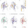
Fig. 2. The PAS domain fold
A. The three-dimensional structure of the PAS A domain of Azotobacter vinelandii NifL (2GJ3) shows the canonical PAS fold with secondary structure elements Aβ to Iβ. A flavin adenine dinucleotide cofactor is bound in a cleft formed by the β-sheet and helices Eα and Fα. An N-terminal flanking α-helix is shown in white. B. Topology diagram of 2GJ3. β-strands are arranged in the order 2-1-5-4-3. C. Residues involved in cofactor binding in 11 different PAS domains mapped onto the structure from A. Color indicates number of structures in which a given residue forms a ligand contact. D. Residues forming intra- or intermolecular contacts to N- or C-terminal flanking α-helices. 34 PAS structures were analyzed and the color code indicates the number of times a certain residue makes a contact. Closely similar results are obtained when only intramolecular contacts to flanking helices are considered. E. Residues involved in dimerization of 26 different PAS domains mapped onto the structure from A. Color code indicates the number of structures in which a given residue contributes to forming the dimer interface.
Fig. 3
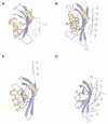
Fig. 3. Diversity of PAS domain structures
Three-dimensional structures of the PAS domains of (A) H. halophila PYP (1MWZ), (B) A. sativa phototropin 1 (2V0U), (C) B. japonicum FixL (1XJ3) and (D) K. pneumoniae CitA (2J80). Secondary structure elements are colored as in Fig. 2A. Other PAS domain structures are shown in Suppl. Fig. 3.
Fig. 4
Fig. 4. Structure-based multiple sequence alignment of PAS domains
Sequences of PAS domains were aligned with respect to their three-dimensional structures and are indicated by their PDB identifiers (Table 1). α-helices and β-sheets are marked by brown and blue shading. Secondary structure elements within the PAS core are labeled. Residues shown in grey italic were not resolved in the structures.
Fig. 5
Fig. 5. Helical domain linkers in PAS-GGDEF proteins
A. Multiple sequence alignment of the linker region between PAS and GGDEF domains. 12 out of 2074 sequences are shown and labeled with their Uniprot identifiers. Residues conserved in more than 50 % of all 2074 sequences are highlighted in bold red, positions with more than 50 % hydrophobic residues by brown shading. Plots below the alignment indicate average sequence conservation and hydropathy. Hydrophobic positions are labeled a and d according to coiled-coil nomenclature (McLachlan and Stewart, 1975). B. Length distribution of linkers between PAS and GGDEF domains. Lengths were determined according to the alignment as the number of residues between the indicated positions (blue arrows). C. Modulo 7 of the distribution shown in B. 94 % of all sequences fall into the length class 7n + 4.
Fig. 6
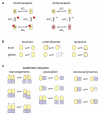
Fig. 6. Signaling by PAS domains
A. Thermodynamic cycle for signal transduction by PAS domains. A protein is in equilibrium between states T and R which differ in biological activity. Presence of a signal alters the free energies of states T and R, and thus shifts the equilibrium between them. Depending on the sensor domain, signal can correspond to binding of a ligand, absorption of a photon, or changes in redox potential or electrical field. B. Models for signal transduction within PAS domains. Signal may induce local or global changes in structure and dynamics in the PAS domain. C. Models for signal propagation to effector domains (blue squares). The activity of oligomeric effector proteins is frequently regulated by quaternary structural changes. In addition, regulation may depend on signal-induced structural and dynamic changes within the effector domain.

References

    1. Alexander RP, Zhulin IB. Evolutionary genomics reveals conserved structural determinants of signaling and adaptation in microbial chemoreceptors. Proc Natl Acad Sci U S A. 2007;104:2885–2890. - PMC - PubMed
    1. Altschul SF, Madden TL, Schaffer AA, Zhang J, Zhang Z, Miller W, Lipman DJ. Gapped BLAST and PSI-BLAST: a new generation of protein database search programs. Nucleic Acids Res. 1997;25:3389–3402. - PMC - PubMed
    1. Anantharaman V, Aravind L. Cache - a signaling domain common to animal Ca(2+)-channel subunits and a class of prokaryotic chemotaxis receptors. Trends Biochem Sci. 2000;25:535–537. - PubMed
    1. Anantharaman V, Balaji S, Aravind L. The signaling helix: a common functional theme in diverse signaling proteins. Biol Direct. 2006;1:25. - PMC - PubMed
    1. Anantharaman V, Koonin EV, Aravind L. Regulatory potential, phyletic distribution and evolution of ancient, intracellular small-molecule-binding domains. J Mol Biol. 2001;307:1271–1292. - PubMed

Publication types

Substances

LinkOut - more resources