Review
. 2009 Oct 14;17(10):1295-306.
doi: 10.1016/j.str.2009.09.001.

Discovery through the computational microscope

Affiliations
Review

Discovery through the computational microscope

Eric H Lee et al. Structure. .

Abstract

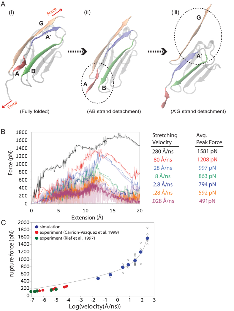
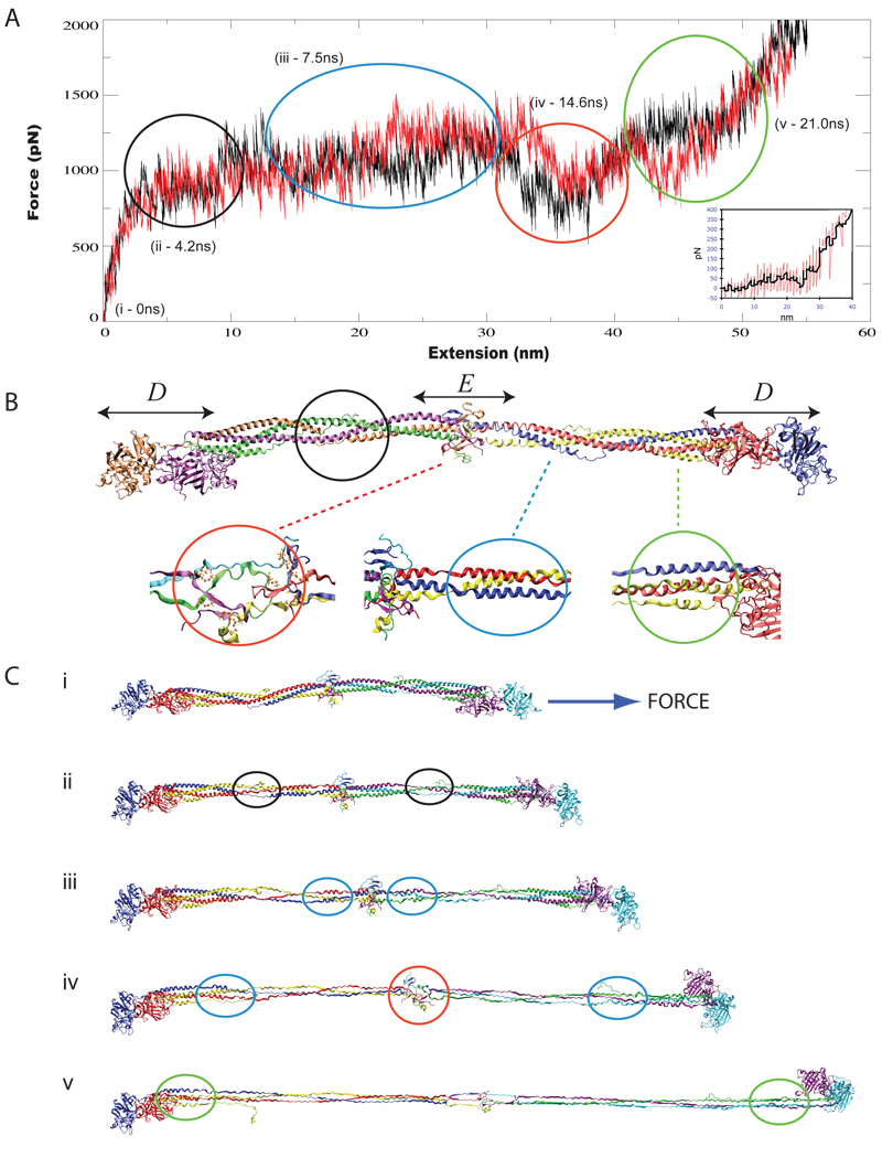
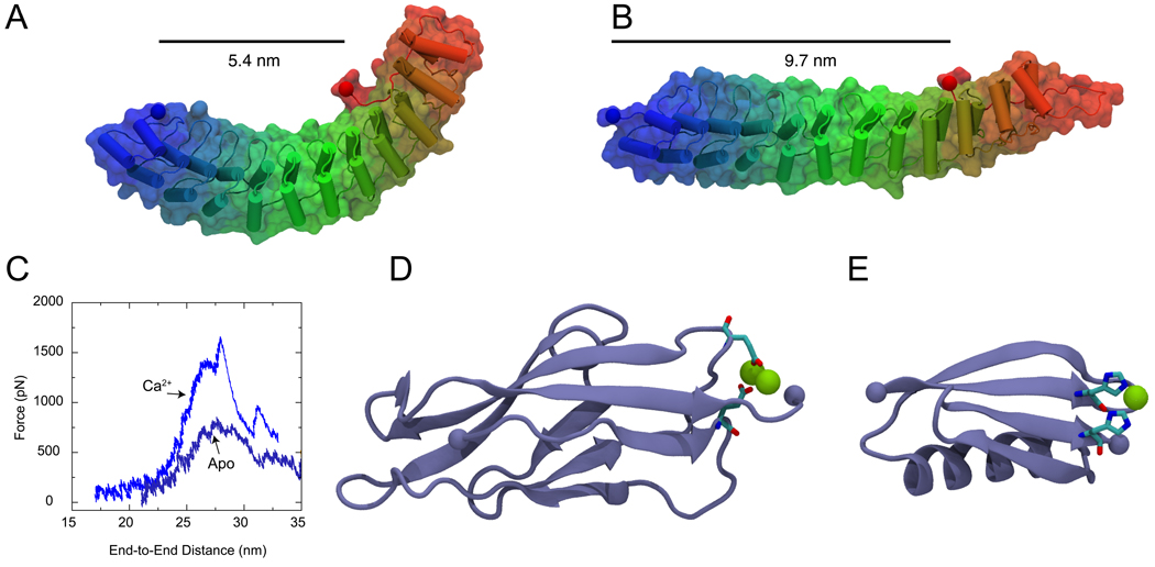
All-atom molecular dynamics simulations have become increasingly popular as a tool to investigate protein function and dynamics. However, researchers are concerned about the short time scales covered by simulations, the apparent impossibility to model large and integral biomolecular systems, and the actual predictive power of the molecular dynamics methodology. Here we review simulations that were in the past both hotly disputed and considered key successes, namely of proteins with mainly mechanical functions (titin, fibrinogen, ankyrin, and cadherin). The simulation work covered shows how state-of-the-art modeling alleviates some of the prior concerns and how unrefuted discoveries are made through the "computational microscope."


Figures

Figure 1
Figure 1
Steered molecular dynamics simulations for stretching fibrinogen. The force-extension curves derived from SMD simulations stretching a whole fibrinogen molecule (1,000,000 atom system) are shown in (A). The black trace is from the SMD simulation reported in (Lim et al., 2008) and the red trace is from a new simulation reported here. An AFM force-extension trace for a single fibrinogen molecule is shown in the inset (from Lim et al. (2008)). Shown in (B) is a cartoon representation of the most complete atomic structure of fibrinogen resolved to date (pdb code 1m1j), showing the distinct terminal D β-sheet globular domains encapsulating mirror-symmetric coiled-coil domains and a central domain, labelled E. SMD simulations stretching the coiled-coil domain of fibrinogen reveal a stepwise unraveling according to helical density. The regions of distinct helical density are also shown in (B), comprised of a two helix region (black circle), a three helix region (blue circle), and a four helix region (green circle). The central domain is denoted by a red circle. Snapshots of the fibrinogen molecule during simulation at time intervals (denoted (i) through (v), also shown in (B)) corresponding to the unraveling of specific regions are shown in (C).
Figure 2
Figure 2
Steered molecular dynamics simulations for stretching fibrinogen. The force-extension curves derived from SMD simulations stretching a whole fibrinogen molecule (1,000,000 atom system) are shown in (A). The black trace is from the SMD simulation reported in (Lim et al., 2008) and the red trace is from a new simulation reported here. An AFM force-extension trace for a single fibrinogen molecule is shown in the inset (from Lim et al. (2008)). Shown in (B) is a cartoon representation of the most complete atomic structure of fibrinogen resolved to date (pdb code 1m1j (Yang et al., 2001)), showing the distinct terminal D β-sheet globular domains encapsulating mirror-symmetric coiled-coil domains and a central domain, labelled E. SMD simulations stretching the coiled-coil domain of fibrinogen reveal a stepwise unraveling according to helical density. The regions of distinct helical density are also shown in (B), comprised of a two helix region (black circle), a three helix region (blue circle), and a four helix region (green circle). The central domain is denoted by a red circle. Snapshots of the fibrinogen molecule during simulation at time intervals (denoted (i) through (v), also shown in (A)), corresponding to the unraveling of specific fibrinogen regions, are shown in (C).
Figure 3
Figure 3
Tertiary structure elasticity of Ankyrin-R and divalent-controlled secondary structure elasticity of cadherin and GB1 proteins. (A) Snapshot of an Ankyrin-R domain in its crystallographic conformation (Michaely et al., 2002). The elongated and curved shape results from parallel stacking of 12 ankyrin repeats. The protein is shown in cartoon and surface representation. (B) Stretched conformation of Ankyrin-R obtained through SMD simulations (Sotomayor et al., 2005). The protein reversibly changes its shape without modifying its secondary structure elements. The end-to-end distance measured from terminal Cα atoms (taking into account the spectrin binding domain) is shown before and after stretching. (C) The unfolding force as a function of distance is shown for C-cadherin simulations performed in the presence (Ca2+) and absence (Apo) of calcium. A significant reduction in the force required to unfold cadherin repeats is observed in the absence of calcium. (D) Snapshot of the first C-cadherin repeat structure (Boggon et al., 2002) shown in cartoon representation. Calcium ions (shown in green) link β-strands A and F through highly conserved and charged amino-acids (shown in licorice representation). (E) Structure of GB1 protein illustrating the position of an engineered histidine-metal chelation site at position G8-55, based on (Cao et al., 2008). (F) Topology of cadherin repeats. A link between β-strands A and F is formed in the presence of Ca2+. (G) Similarly, a link between GB1 β-strands A and D is formed upon addition of Ni2+. The links enhance the mechanical stability of these proteins.

References

    1. Adcock SA, McCammon JA. Molecular dynamics: survey of methods for simulating the activity of proteins. Chem. Rev. 2006;106:1589–1615. - PMC - PubMed
    1. Aksimentiev A, Brunner R, Cohen J, Comer J, Cruz-Chu E, Hardy D, Rajan A, Shih A, Sigalov G, Yin Y, et al. Protocols in Nanostructure Design. Methods in Molecular Biology. Humana Press; 2008. Computer modeling in biotechnology, a partner in development; pp. 181–234. - PubMed
    1. Arkhipov A, Freddolino PL, Schulten K. Stability and dynamics of virus capsids described by coarse-grained modeling. Structure. 2006;14:1767–1777. - PubMed
    1. Arkin IT, Xu H, Jensen M, Arbely E, Bennett ER, Bowers KJ, Chow E, Dror RO, Eastwood MP, Flitman-Tene R, et al. Mechanism of Na+/H+ antiporting. Science. 2007;317:799–803. - PubMed
    1. Bang ML, Centner T, Fornoff F, Geach AJ, Gotthardt M, McNabb M, Witt CC, Labeit D, Gregorio CC, Granzier H, et al. The complete gene sequence of titin, expression of an unusual approximate to 700-kDa titin isoform, and its interaction with obscurin identify a novel Z-line to I-band linking system. Circulation Res. 2001;89:1065–1072. - PubMed

Publication types