. 2009 Oct 14;17(10):1377-85.
doi: 10.1016/j.str.2009.08.010.

Structural insights on the Mycobacterium tuberculosis proteasomal ATPase Mpa

Affiliations

Structural insights on the Mycobacterium tuberculosis proteasomal ATPase Mpa

Tao Wang et al. Structure. .

Abstract

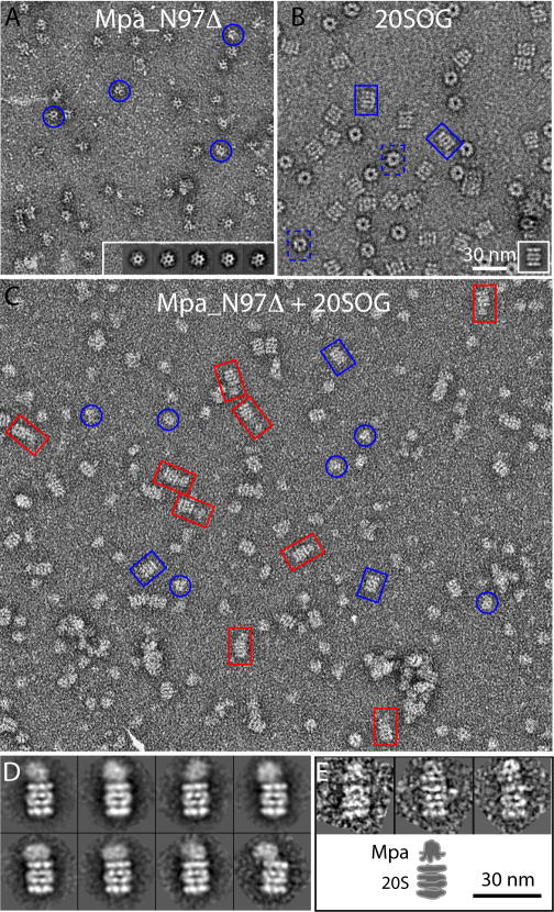
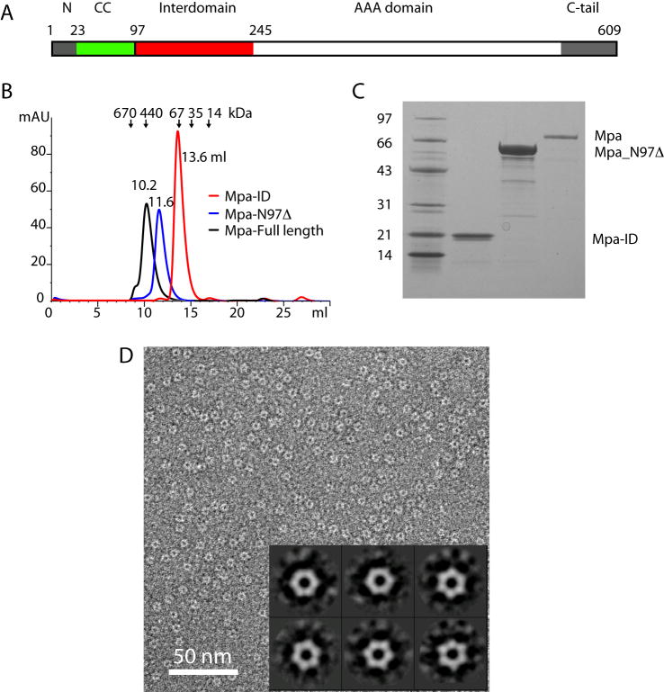
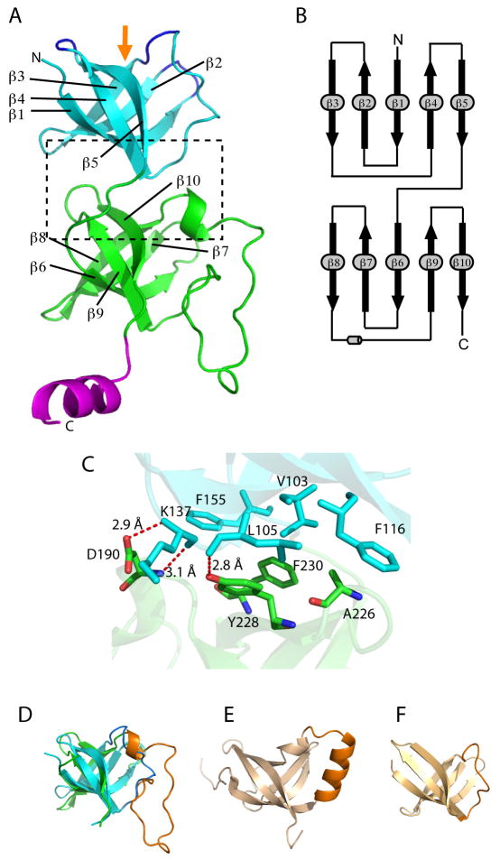
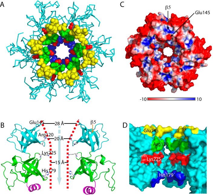
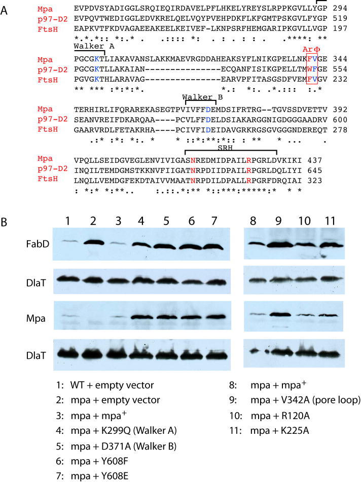
Proteasome-mediated protein turnover in all domains of life is an energy-dependent process that requires ATPase activity. Mycobacterium tuberculosis (Mtb) was recently shown to possess a ubiquitin-like proteasome pathway that plays an essential role in Mtb resistance to killing by products of host macrophages. Here we report our structural and biochemical investigation of Mpa, the presumptive Mtb proteasomal ATPase. We demonstrate that Mpa binds to the Mtb proteasome in the presence of ATPgammaS, providing the physical evidence that Mpa is the proteasomal ATPase. X-ray crystallographic determination of the conserved interdomain showed a five stranded double beta barrel structure containing a Greek key motif. Structure and mutational analysis indicate a major role of the interdomain for Mpa hexamerization. Our mutational and functional studies further suggest that the central channel in the Mpa hexamer is involved in protein substrate translocation and degradation. These studies provide insights into how a bacterial proteasomal ATPase interacts with and facilitates protein degradation by the proteasome.


Figures

Fig. 1
Fig. 1
Mpa directly interacts with the open gate form (20SOG) of the Mtb proteasome. (A) A small region of a raw electron micrograph of negatively stained Mpa_N97Δ. Inset: Five class averages of the deletion mutant Mpa particles. The blue circles mark several particles. (B) A raw EM image of the Mtb open gate 20S proteasome. Inset: 2D class average of the proteasome particles in the side view. The blue rectangles mark individual particles in side (solid line) or top (dashed line) views. (C) A raw EM micrograph of 20SOG incubated with Mpa_N97Δ in the presence of ATPγS. The red rectangles mark several 20CP-Mpa complexes. Panels (A) and (B) are in the same scale. (D) Eight selected 2D class averages of the Mpa-20SOG complex particles showing the flexible nature of the interaction between Mpa and proteasome. (E) Upper panel: three raw particle images of the Mpa-20SOG complex in which the Mpa binds approximately axially on the proteasome. Lower panel: a sketch showing that the Mpa C-terminal portion with weaker density interfaces with the 20SOG. Panels (D) and (E) are in the same scale. Scale bar: 30 nm.
Fig. 2
Fig. 2
Mpa has a multi-domain architecture. (A) Overview of the domain organization of the full-length Mpa. (B) The Superdex-200 gel filtration profiles of the purified full-length Mpa in black, Mpa_N97Δ in blue, and Mpa-ID in red. Each of the three proteins is eluted at the volume corresponding to their respective hexameric form. (C) The Coomassie blue stained SDS-PAGE of the purified Mpa-ID (lane 2), Mpa_N97Δ (lane 3), and Mpa (lane 4). Lane 1 is the molecular weight marker in kD. (D) The negatively stain EM image of the purified Mpa-ID and the inserted six 2D class averages reveal a hexameric structure about 65 Å in diameter with a ~20 Å central low density channel.
Fig. 3
Fig. 3
The crystal structure of the Mpa-ID. (A) Mpa-ID contains two five-stranded β-barrel subdomains connected by a loop. The C-terminal helix might be a linker between the interdomain and the following AAA domain. The orange arrow indicates the substrate recognition/binding region at the top as defined by the three β-hairpin loops highlighted in blue. (B) The topology of Mpa-ID contains two Greek key motifs formed by the first four β-strands in each sub-domain. (C) An enlarged view of the interface between the two tandem β-barrels as boxed in (A), showing the hydrophobic and H-bond interactions. (D) The two β-barrel sub-domains have a similar fold; they can be overlapped with an RMSD of 1.9Å. (E) and (F) are the OB-fold structures of the vaccinia virus K3L (D, PDB ID 1LUZ, RMSD = 2.5 Å) and the transcription factor 5A (E, PDB ID 1EIF, RMSD = 1.5 Å, sequence identity = 21%), respectively, aligned with the first β-barrel sub-domain in Mpa-ID.
Fig. 4
Fig. 4
Crystal structure of the Mpa-ID hexamer. (A) The top view of the hexamer, with residues of the four β-hairpins lining the central channel shown as spheres in different colors. (B) Side view showing only two diagonal subunits. The four residues lining the funnel-like central channel are highlighted in the ball-and-stick presentation. The potential peptide translocation path is illustrated by a cyan arrow. (C) Surface-charge presentation of the Mpa-ID hexamer in the top view. Arrows indicate the substrate-binding site in the OB-fold above the β5 strand, and the negatively charged Glu-145 at the opening of the channel, respectively. (D) Surface rendered side view of the Mpa-ID hexamer with the front two subunits removed to show the interior channel. Charged residues lining the channels are labeled and shown in different colors.
Fig. 5
Fig. 5
Site-directed mutations in Mpa disrupt protein degradation. (A) Sequence alignment of Mpa with p97/VCP and FtsH reveals unexpected high identity at the α/β sub-domain of the AAA domain with p97 D2 domain at 46% and FtsH at 38.5%. The conserved aromatic and hydrophobic residues in the pore-loop region of AAA domain are highlighted by a red box (ArΦ). The Walker A/B motifs, and the conserved sensor-1 residue (N) and the arginine finger (R) in the second region of homology (SRH) are also marked. (B) Immunoblot analysis of total Mtb lysates of equivalent cell numbers of wild type, mpa mutant or mpa mutant Mtb transformed with plasmids encoding wild type or mutated mpa. Samples were analyzed with either polyclonal antibodies to FabD or Mpa. The same blots were stripped and re-analyzed with antibodies to dihydrolipoamide dehydrogenase (DlaT), the loading control.
Fig. 6
Fig. 6
Subunit-subunit interactions in the Mpa-ID hexamer. (A) The interface between two neighboring β-barrel one is limited, involving only two main chain H-bonds between the Arg-123 and Ser-99. (B) The interactions between the two neighboring β-barrel two are extensive. At the top of the interface, Trp-187 and Val-158 form a hydrophobic interaction; at the middle section, the interface involves two salt bridges/H-bonds (between Arg-173 and Glu-231, and between Glu-183 and Lys-235), and a pair of hydrophobic residues (between Ile-233 and Leu-168); and at the bottom section, Arg-165 and Glu-166 form two H-bonds with the Ala-236. (C). Gel filtration profiles of the native full-length Mpa, Mpa_N97Δ, the full-length Mpa triple mutant (Mpa‴), Mpa-ID, and Mpa-ID with the triple mutations (Mpa-ID‴). (D) The Blue Native gel of the corresponding Mpa constructs purified as shown in (C). The right panel (lane 7 and 8) was run separately for a shorter period of time than the left panel. This was to prevent the small Mpa-ID‴ (~ 20 kD) from running out of the gel.

Comment in

  • How ATPases unravel a mystery.
    Gallastegui N, Groll M. Gallastegui N, et al. Structure. 2009 Oct 14;17(10):1279-81. doi: 10.1016/j.str.2009.09.003. Structure. 2009. PMID: 19836328

References

    1. Alexandrescu AT, Gittis AG, Abeygunawardana C, Shortle D. NMR structure of a stable “OB-fold” sub-domain isolated from staphylococcal nuclease. J Mol Biol. 1995;250:134–143. - PubMed
    1. Alexandrescu AT, Jaravine VA, Dames SA, Lamour FP. NMR hydrogen exchange of the OB-fold protein LysN as a function of denaturant: the most conserved elements of structure are the most stable to unfolding. J Mol Biol. 1999;289:1041–1054. - PubMed
    1. Baker TA, Sauer RT. ATP-dependent proteases of bacteria: recognition logic and operating principles. Trends Biochem Sci. 2006;31:647–653. - PMC - PubMed
    1. Benaroudj N, Zwickl P, Seemuller E, Baumeister W, Goldberg AL. ATP hydrolysis by the proteasome regulatory complex PAN serves multiple functions in protein degradation. Mol Cell. 2003;11:69–78. - PubMed
    1. Borissenko L, Groll M. 20S proteasome and its inhibitors: crystallographic knowledge for drug development. Chem Rev. 2007;107:687–717. - PubMed

Publication types

MeSH terms

Substances

Associated data

LinkOut - more resources