. 2010 Jan;90(1):155-67.
doi: 10.1016/j.exer.2009.10.004. Epub 2009 Oct 15.

Low nitric oxide synthases (NOSs) in eyes with age-related macular degeneration (AMD)

Affiliations

Low nitric oxide synthases (NOSs) in eyes with age-related macular degeneration (AMD)

Imran A Bhutto et al. Exp Eye Res. 2010 Jan.

Abstract

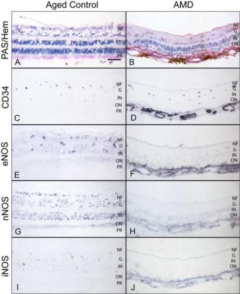
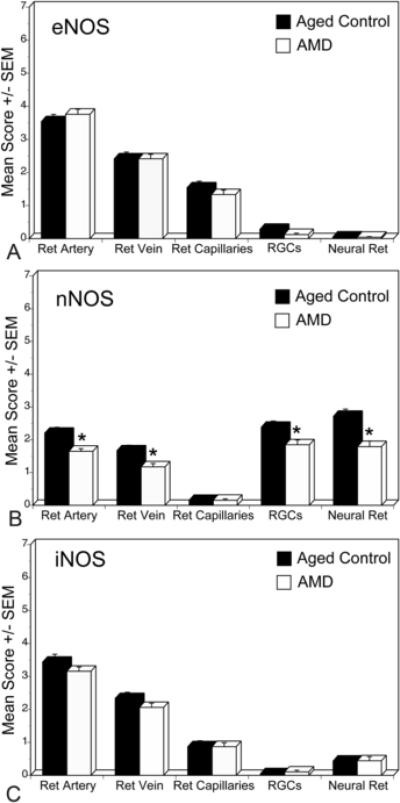
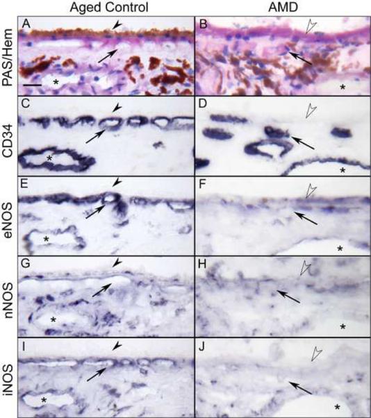
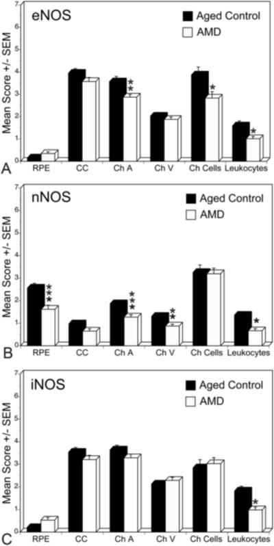
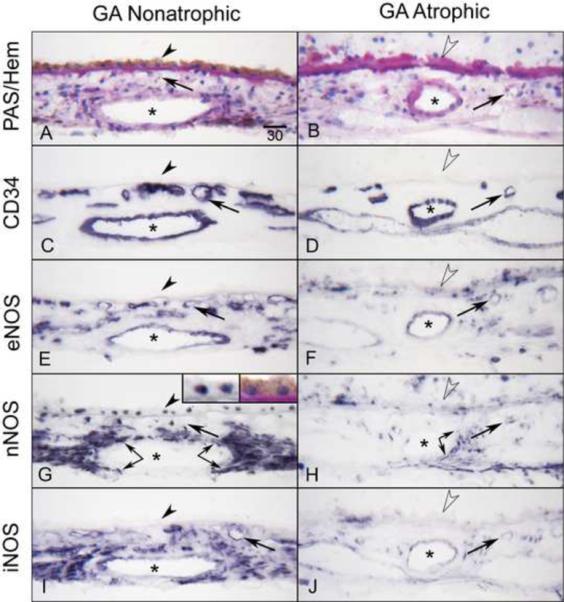
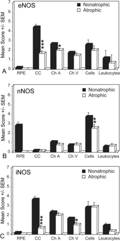
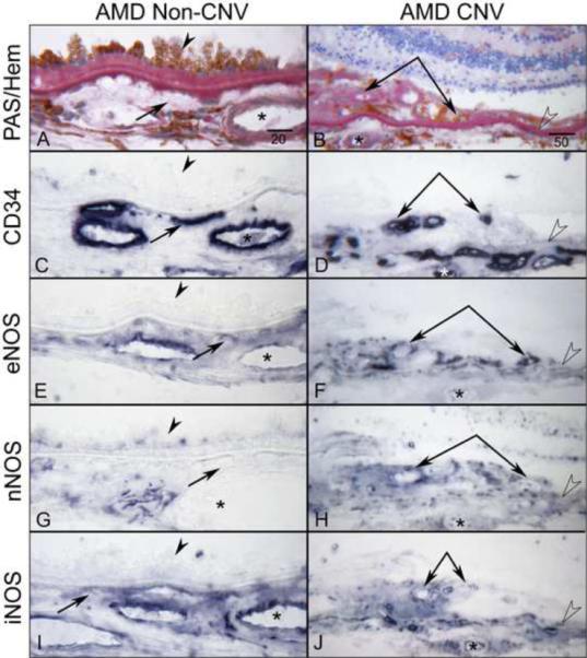
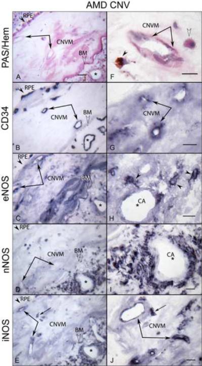
Nitric oxide (NO) production by vascular endothelium is important in regulation of blood flow. Reduced production of NO can adversely affect blood flow and other vascular functions. We investigated the expression of three nitric oxide synthase (NOS) isoforms in retina and choroid of aged human eyes and eyes with AMD. Alkaline phosphatase immunohistochemistry was performed using antibodies against inducible (iNOS), neuronal (nNOS), and endothelial (eNOS) NOSs on cryopreserved sections from aged control donor eyes (n = 13) and eyes with AMD (n = 22). CD34 antibody was used as an endothelial cell (EC) marker. Three independent masked observers scored the intensity of the immunohistochemical reaction product. Mean scores from the aged control and AMD eyes were statistically compared. In aged control retinas, nNOS was in ganglion cells (RGCs) and neurons of both nuclear layers. In choroid, perivascular nerve fibers and retinal pigment epithelial (RPE) cells were nNOS+. eNOS and iNOS were confined to the retinal and choroidal vascular ECs. Some cells presumably melanocytes or dendritic cells in choroid were also eNOS+. In AMD eyes, nNOS was significantly lower in RGCs, neurons, retinal vessels and RPE (p < or = 0.05) compared to the aged control eyes. iNOS and eNOS showed no significant differences between aged control and AMD eyes except that there was significantly less eNOS in choroidal arteries (p = 0.006) and choroidal cells (p = 0.03) of AMD eyes. Although NO was not measured directly, these findings suggest that there is less NO produced in AMD eyes. The decrease in retinal nNOS in AMD eyes is probably related to neuronal degeneration. The decrease in nNOS and eNOS in AMD choroid could be associated with vasoconstriction and hemodynamic changes.


Figures

Figure 1
Figure 1
Immunoreactivity for NOS isoforms in the aged control (subject 10) and AMD (subject 18) retina. PAS and hematoxylin staining shows normal morphological features of the aged control (A) and a degenerative thin retina with loss of photoreceptors in AMD (B). Retinal blood vessels are labeled with CD34 (C, D). Note the AMD retina is thin so choroidal blood vessels are present in the pictures and stain with CD34 (D). In aged control retina, eNOS antibody staining is present in the retinal blood vessels and in a few scattered cells in ganglion cell and inner nuclear layer, which may be retinal capillaries (E). nNOS is prominent in ganglion cells and neurons in both inner and outer nuclear layers (G). iNOS is present in a few scattered cells in the inner nuclear layer (I). In this AMD retina, immunoreactivity for NOS isoforms is significantly weaker than in the control retina (F–J). Magnification bar (A–J) = 100 μm. (NF=nerve fiber layer; G=ganglion cell layer; IN=Inner nuclear; ON=outer nuclear; PR=photoreceptors)
Figure 2
Figure 2
Mean immunoreactivity scores±SEM for NOS isoforms in retinal structures of all aged control (black) and AMD (white) eyes. The immunoreactivity scores for nNOS (B) were significantly lower in RGCs, neural cells, and retinal arteries and veins in AMD retina compared to aged control. There were no significant difference in eNOS (A) and iNOS (C) immunoreactivity levels for retinal structures between aged and AMD retinas. The significance of the difference between the groups by t test is indicated: *= p<0.05.
Figure 3
Figure 3
Immunoreactivity for NOS isoforms in aged control (subject 2) and AMD (subject 13) choroid. PAS and hematoxylin staining show morphological features of choroids (A, B). Note that thickened PAS positive Bruch's membrane (open arrowheads) in AMD choroid (B). In aged control choroid, large and medium choroidal vessels (asterisk) and choriocapillaris (CC; arrows) are intensely labeled for CD34 and appear morphologically normal with broad lumens (C), whereas there is loss of CC and CC lumens appear constricted in AMD choroid (D). In aged control choroid, eNOS is prominently localized to the CC and, to a lesser extent, in endothelial cells of medium sized choroidal blood vessels and a few individual cells in choroidal stroma (E). nNOS is present in RPE nuclei, and perivascular nerve fibers and cells in stroma (G). iNOS is localized to endothelial cells of blood vessels and individual cells in stroma (I). In AMD choroid, immunoreactivity for NOS isoforms is greatly reduced compared to the control subject (F, H, J). Magnification bar (A–J) = 20 μm.
Figure 4
Figure 4
Mean immunoreactivity scores±SEM for NOS isoforms in choroidal structures of aged control (black) and AMD (white) eyes. The immunoreactivity scores for eNOS were significantly lower in choroidal arteries, cells in stroma, and leukocytes in blood vessel lumens in AMD choroid. nNOS was significantly lower in RPE nuclei, arteries and veins as well as leukocytes. There was no significant difference in iNOS levels between aged control and AMD choroids except in leukocytes. The significance of the difference between the groups by t test is indicated: *= p<0.01, **= p<0.005, ***= p<0.001.
Figure 5
Figure 5
Immunoreactivity for NOS isoforms in AMD eye with geographic atrophy (subject 15). Note that the non-atrophic area (A) of choroid has RPE (arrowhead) and there is PAS-positive thickened Bruch's membrane (open arrowhead) but there is no RPE in atrophic area (B). CD34 demonstrates CC (arrows) is limited in the atrophic area compared to the non-atrophic area (C, D). In non-atrophic area, eNOS (E) is prominent in CC and endothelial cells of large blood vessels (asterisks) as well as cells in stroma, which may include melanocytes. nNOS (G) is prominent in RPE nuclei (at high magnification, Inset) and perivascular nerve fibers (double arrows). iNOS (I) appears similar to nNOS. The similarity with nNOS may be due to cross reactivity of the antibodies. Immunoreactivity for NOS isoforms is greatly reduced in choroid structures in the atrophic area (F, H, J). Magnification bar (A–J) = 30 μm.
Figure 6
Figure 6
Mean immunoreactivity scores±SEM for NOS isoforms in choroidal structures comparing atrophic (black) and non-atrophic (white) areas in a geographic atrophy (GA) subjects. The immunoreactivity scores for eNOS were significantly lower in choroidal arteries and CC in atrophic area compared to the non-atrophic area. The mean score for nNOS was significantly lower in perivascular nerve fibers, whereas the score for iNOS was significantly lower in choroidal veins and CC in atrophic area than in the non-atrophic area. The significance of the difference between the groups by t test is indicated: *= p<0.01, **= p<0.005, ***= p<0.001.
Figure 7
Figure 7
Immunoreactivity for NOS isoforms in subretinal CNV and choroid under and adjacent to CNV (non-CNV) areas in late AMD eye (subject 19). PAS and hematoxylin staining demonstrates basal laminar deposits under hypertrophic RPE (arrowhead) in choroid with no CNV (A) and subretinal CNV (double arrows) with PAS-positive Bruch's membrane (open arrowhead) (B). The CC (arrows), choroidal vessels (asterisks) and subretinal CNV (double arrows) are intensely positive for CD34 (C, D). Note the moderate immunoreactivity for all three NOS isoforms is seen in subretinal CNV (F, H, J), whereas weak staining for nNOS is observed not only in choroid underneath the subretinal CNV but in the choroid adjacent to CNV (G, H). Moderate eNOS and iNOS staining are observed in choroid with no CNV (E, I). Magnification bar in left panels = 20 μm and in right panels = 50 μm.
Figure 8
Figure 8
Immunoreactivity for NOS isoforms in CNV within disciform scar and the choroid underneath the scar in late AMD eye (subject 20). In left panels, (A) PAS and hematoxylin staining demonstrates CNV within scar (double arrows) and PAS-positive Bruch's membrane (BM; open arrowhead) and choroidal vessels (asterisks) underneath the scar. RPE are indicated by arrowhead. (B) The CNV and choroidal vessels are positive for CD34. Immunoreactivity for eNOS (C) and iNOS (E) is prominent in CNV, whereas the nNOS is negative in CNV (D). Note that the choroidal cells and perivascular nerve fibers of choroid underneath the scar exhibit intense immunoreactivity for NOS isoforms (C, D, E). In right panels (at higher magnification), PAS-positive pigmented cells (arrowhead) and apparent monocytes (open arrowhead) are seen close to CNV (double arrows) (F). The CNV is positive for CD34 (G). The intense eNOS immunoreactivity in single cells presumably monocytes in choroid underneath the scar (H), whereas nNOS is present in perivascular nerve fibers (I). (J) iNOS is in CNV and presumably monocytes (arrow). Magnification bar in left panels = 50 μm and right panels = 20 μm.

References

    1. Alderton WK, Cooper CE, Knowles RG. Nitric oxide synthases: structure, function and inhibition. Biochem J. 2001;357:593–615. - PMC - PubMed
    1. Beatty S, Koh H, Phil M, Henson D, Boulton M. The role of oxidative stress in the pathogenesis of age-related macular degeneration. Surv Ophthalmol. 2000;45:115–134. - PubMed
    1. Bergua A, Mayer B, Neuhuber WL. Nitrergic and VIPergic neurons in the choroid and ciliary ganglion of the duck Anis carina. Anat Embryol (Berl) 1996;193:239–248. - PubMed
    1. Bernatchez PN, Bauer PM, Yu J, Prendergast JS, He P, Sessa WC. Dissecting the molecular control of endothelial NO synthase by caveolin-1 using cell-permeable peptides. Proc Natl Acad Sci U S A. 2005;102:761–766. - PMC - PubMed
    1. Bhutto IA, Kim SY, McLeod D, S, Merges CA, Fukai N, Olsen BR, Lutty GA. Localization of collagen XVIII and the endostatin portion of collagen XVIII in aged human control eyes and eyes with age-related macular degeneration. Invest. Ophthalmol. Vis. Sci. 2004;45:1544–1552. - PubMed

Publication types