Review
. 2010 Jan;31(1):32-43.
doi: 10.1016/j.yfrne.2009.10.006. Epub 2009 Oct 24.

Sweet talk in the brain: glucosensing, neural networks, and hypoglycemic counterregulation

Affiliations
Review

Sweet talk in the brain: glucosensing, neural networks, and hypoglycemic counterregulation

Alan G Watts et al. Front Neuroendocrinol. 2010 Jan.

Abstract

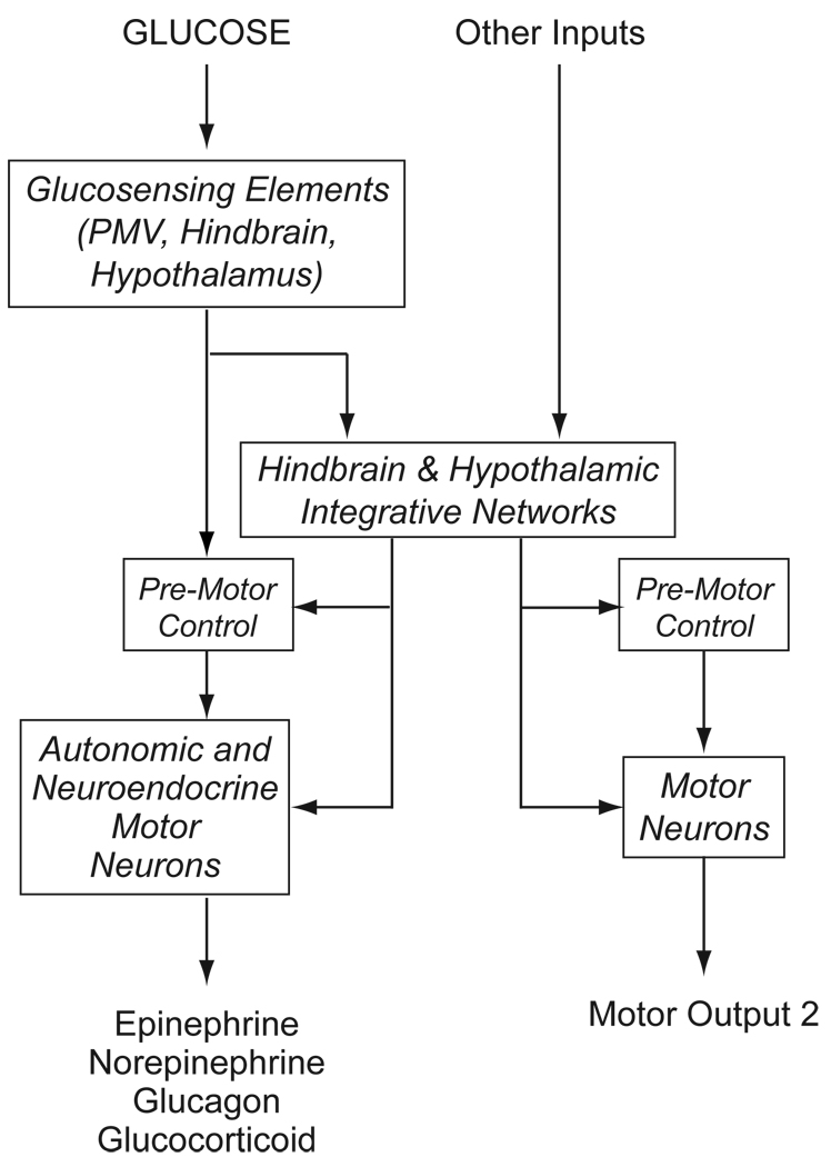
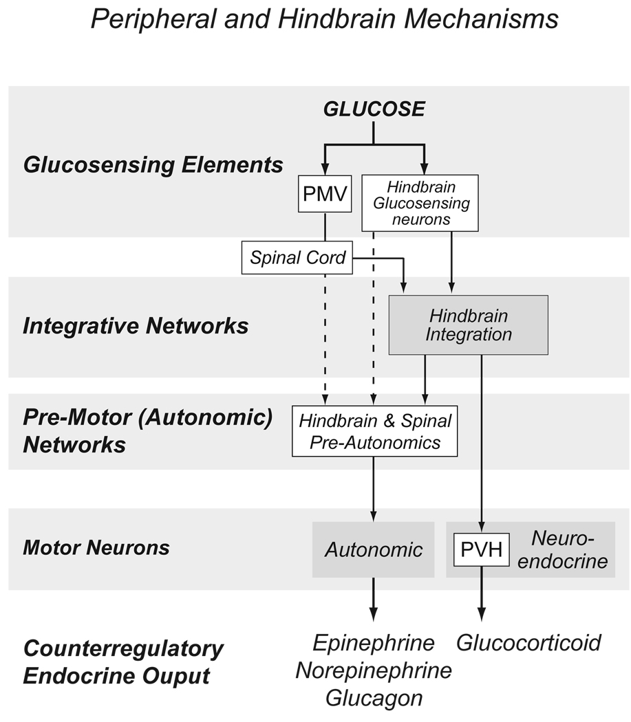
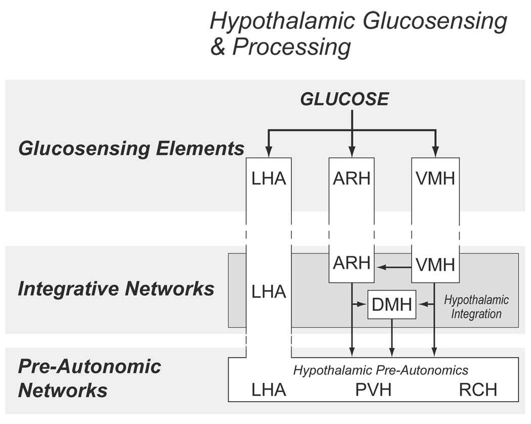
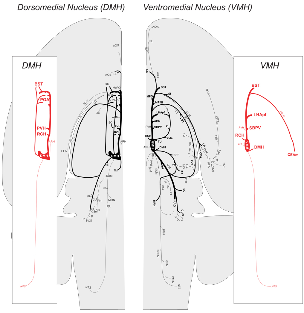
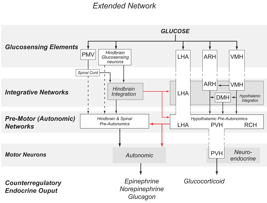
Glucose is the primary fuel for the vast majority of cells, and animals have evolved essential cellular, autonomic, endocrine, and behavioral measures to counteract both hypo- and hyperglycemia. A central component of these counterregulatory mechanisms is the ability of specific sensory elements to detect changes in blood glucose and then use that information to produce appropriate counterregulatory responses. Here we focus on the organization of the neural systems that are engaged by glucosensing mechanisms when blood glucose concentrations fall to levels that pose a physiological threat. We employ a classic sensory-motor integrative schema to describe the peripheral, hindbrain, and hypothalamic components that make up counterregulatory mechanisms in the brain. We propose that models previously developed to describe how the forebrain modulates autonomic reflex loops in the hindbrain offer a reasoned framework for explaining how counterregulatory neural mechanisms in the hypothalamus and hindbrain are structured.


Figures

Figure 1
Figure 1
A schematic diagram to illustrate how glucose, acting as an interosensory stimulus engages glucosensing elements that transduce information into neural signals that control hypoglycemic counterregulation. Consistent with classic sensorimotor integration, these signals use neural pathways to engage either premotor control networks to drive autonomic and neuroendocrine motor neurons in a reflex manner; or more complex integrative networks in the hypothalamus and hindbrain. These integrative networks allow glucosensation to be integrated with other information that can modulate counterregulatory responses, as well as providing a way for glucosensation to influence other motor process, such as those that control reproductive behavior.
Figure 2
Figure 2
The magnitude of the ACTH response to hypoglycemia varies across the day. The two panels show the incremental change in rat plasma ACTH concentrations 20mins after an intravenous injection of insulin given 2h after lights on (A, open circles) or 1h before lights off (B, closed circles). Each symbol represents the response of a single animal. Dashed horizontal lines represent 2 SD above the mean increment of the saline-injected controls at that time. The vertical shaded bar in B) denotes the range of glucose concentrations containing the glycemic threshold. (Data from [45]).
Figure 3
Figure 3
An expansion of the model shown in Figure 1 to illustrate the organization of hindbrain and peripheral components involved with counterregulation. Solid lines represent known projections; dotted lines represent postulated connections. Abbreviations: PMV, hepatic portal/mesenteric vein; PVH, paraventricular nucleus of the hypothalamus.
Figure 4
Figure 4
An expansion of the model shown in Figure 1 to illustrate the organization of hypothalamic components involved with counterregulation. Solid lines represent known projections. Abbreviations: ARH, arcuate nucles; DMH, dorsomedial nucleus; LHA, lateral hypothalamic area; PVH, paraventricular nucleus of the hypothalamus; RCH, retrochiasmatic area; VMH, ventromedial nucleus.
Figure 5
Figure 5
The efferent projections of the dorsomedial nucleus (DMH) and ventromedial nucleus (VMH) as revealed by the anterograde tracer Phaseolus vulgaris leucoagglutinin (PHAL) and then plotted onto a flat map of the rat brain [109]. The thickness of the lines represent the relative strengths of each projection. The side panels emphasize those projections from each nucleus to those regions that are most closely associated with metabolic regulation. Note the paucity of projections from each nucleus to those regions of the hindbrain important for autonomic function. (Adapted from [21,115]). Abbreviations in the side panels : ap, ansa peduncularis; ARH, arcuate nucleus hypothalamus; BST, bed nuclei stria terminalis; CEAm, central nucleus amygdala, medial part; DMH, dorsomedial nucleus hypothalamus; LHApf, lateral hypothalamic area, perifornical part; NTS, nucleus ofthe solitary tract; POA, preoptic area; PVH, paraventricular nucleus hypothalamus; RCH, retrochiasmatic area; SBPV, subparavenrticular zone; st, stria terminalis; VMH, ventromedial nucleus hypothalamus. Please refer to [21,115] for other abbreviations.
Figure 6
Figure 6
An expansion of the model shown in Figure 1 to illustrate interactions between the expanded network of peripheral, hindbrain and hypothalamic components involved with hypoglycemic counterregulation. Solid lines represent known projections. Connections between the hypothalamus and hindbrain are shown in red. Abbreviations: as Fig 3 and Fig 4.

Similar articles

Cited by

References

    1. Adachi A, Shimizu N, Oomura Y, Kobáshi M. Convergence of hepatoportal glucose-sensitive afferent signals to glucose-sensitive units within the nucleus of the solitary tract. Neurosci. Letters. 1984;46:215–218. - PubMed
    1. Anand BK, Brobeck JR. Hypothalamic control of food intake in rats and cats. Yale J. Biol. Med. 1951;24:123–140. - PMC - PubMed
    1. Anand BK, Chhina GS, Sharma KN, Dua S, Singh B. Activity of single neurons in the hypothalamic feeding centers: effect of glucose. Am. J. Physiol. 1964;207:1146–1154. - PubMed
    1. Andrew SF, Dinh TT, Ritter S. Localized glucoprivation of hindbrain sites elicits corticosterone and glucagon secretion. Am. J. Physiol. Regul. Integr. Comp. Physiol. 2007;292:R1792–R1798. - PubMed
    1. Balfour RH, Hansen AM, Trapp S. Neuronal responses to transient hypoglycaemia in the dorsal vagal complex of the rat brainstem. J. Physiol. 2006;570:469–484. - PMC - PubMed

Publication types